. 2008 Dec 3;28(49):13056-65.
doi: 10.1523/JNEUROSCI.1307-08.2008.

Short interfering RNA against transient receptor potential vanilloid 1 attenuates cisplatin-induced hearing loss in the rat

Affiliations

Short interfering RNA against transient receptor potential vanilloid 1 attenuates cisplatin-induced hearing loss in the rat

Debashree Mukherjea et al. J Neurosci. .

Abstract

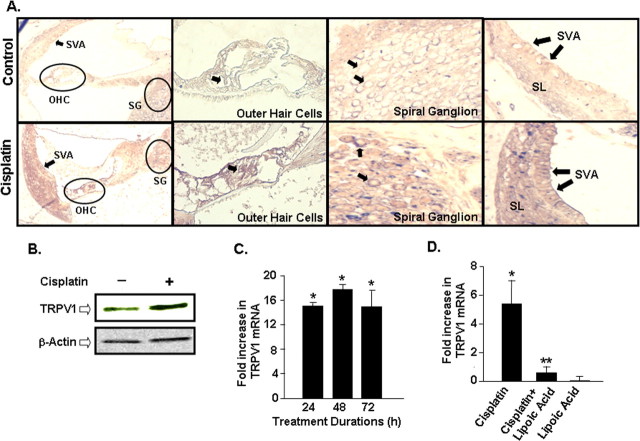
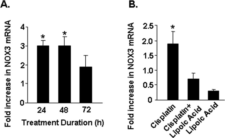
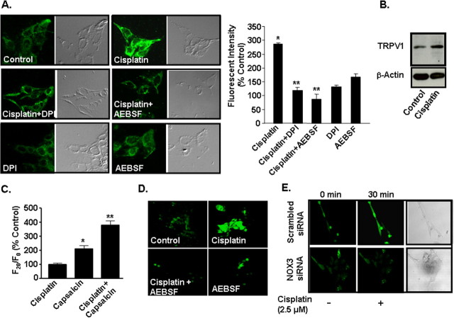
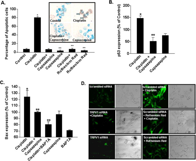
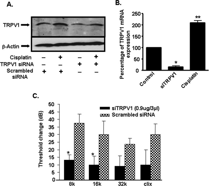
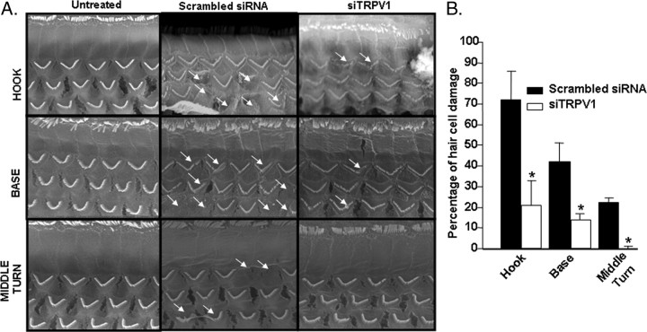
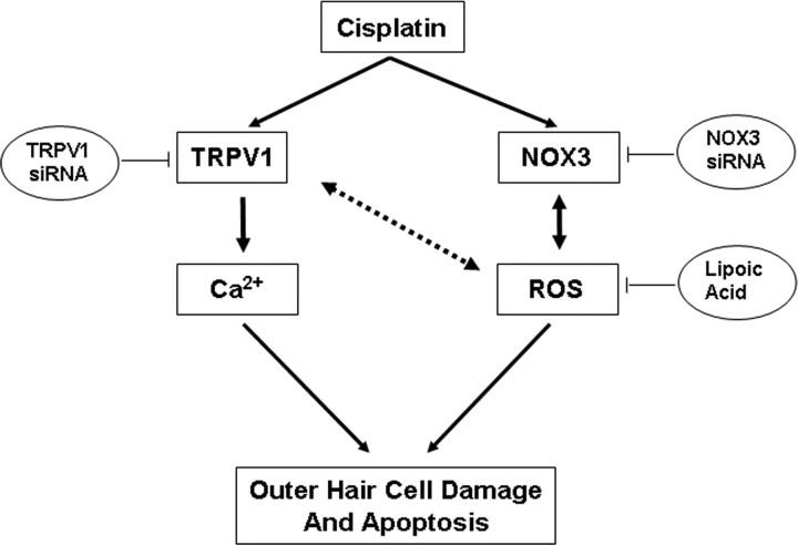
Cisplatin, a chemotherapeutic agent of choice for the treatment of solid tumors, produces hearing loss in approximately half a million new cancer patients annually in the United States. The hearing loss is due, in part, to increased generation of reactive oxygen species (ROS) in the cochlea, leading to lipid peroxidation and damage or death of outer hair cells in the organ of Corti. The cochlea expresses the transient receptor potential vanilloid 1 (TRPV1), which are normally expressed on small diameter neurons in the peripheral nervous system and mediate thermal sensitivity, but whose role in the cochlea is unclear. In this study, we show that TRPV1 is coregulated along with the NADPH oxidase isoform, NOX3, by cisplatin. Induction of these proteins by cisplatin is dependent on ROS generation, since it is reversed by systemic lipoic acid administration. In organ of Corti hair cell cultures (UB/OC-1 cells), cisplatin activates and induces TRPV1 and NOX3, leading to apoptosis of these cells. Inhibition of TRPV1 by capsazepine or ruthenium red reduced the apoptosis, implicating TRPV1 in this process. Treatment of UB/OC-1 cultures with short interfering RNA (siRNA) against either TRPV1 or NOX3 reduced cisplatin-induced apoptosis, while round window application of TRPV1 siRNA to rats reduced TRPV1 expression, decreased damage to outer hair cells and reduced cisplatin-induced hearing loss. These data provide a link between NOX3 and TRPV1 in cisplatin-induced hearing loss and suggest that targeting these proteins for knockdown by siRNA could serve as a novel approach in treating cisplatin ototoxicity.


Figures

Figure 1.
Figure 1.
Increased TRPV1 expression in organ of Corti after cisplatin administration in vivo. A, Rats were administered vehicle or cisplatin (13 mg/kg, i.p.), and cochleae were harvested after 3 d, decalcified in CAL-EX II over 3 weeks, sectioned and stained with a polyclonal antibody for TRPV1 and counterstained with toluidine blue. Increased immunostaining was observed in cisplatin treated samples, compared with vehicle-treated controls, in the outer hair cells (OHC) and spiral ganglion cells (SG) (arrows). Labeling also appears increased in the stria vascularis (SVA) (arrows). SL represents the spiral ligament. Magnification, 100 ×. B, Increased TRPV1 protein expression is seen in whole cochlear homogenates of rats treated with cisplatin for 72 h compared with untreated control cochleae. Western blot shown is the representative of five different experiments with identical results. C, Time course of cisplatin-mediated increase in TRPV1 mRNA in the rat cochlea shows significant increase in TRPV1 mRNA as early as 24 h compared with vehicle-treated control cochleae. The levels of TRPV1 mRNA were determined using real-time PCR. Asterisk indicates statistically significant increase from vehicle-treated controls (p < 0.05). D, Inhibition of cisplatin-induced TRPV1 expression in the rat cochlea by lipoic acid. Rats were pretreated with vehicle or lipoic acid (50 mg/kg, i.p.) before the administration of cisplatin (13 mg/kg, i.p.) and cochleae were obtained 24 h later for the isolation of RNA and real-time PCR. Single asterisk indicates statistically significant increase from control, while double asterisks indicate statistically significant suppression of the cisplatin-response (n = 5, p < 0.05).
Figure 2.
Figure 2.
Cisplatin increased the expression of NADPH oxidase in the cochlea. A, Time course of induction of NOX3 mRNA by cisplatin. Rats were administered cisplatin (13 mg/kg) and were killed at different time points at which their cochleae were dissected out and processed for RNA and real-time PCR for NOX3. B, Lipoic acid (50 mg/kg, i.p.) abolished cisplatin-mediated increase in NOX3 mRNA. Asterisk indicates statistically significant change compared with vehicle-treated controls (p < 0.05, n = 5).
Figure 3.
Figure 3.
Cisplatin increased TRPV1 expression through activation of NADPH oxidase in UB/OC-1 cells. A, UB/OC-1 cells were pretreated with vehicle, DPI (10 μm) or AEBSF (100 μm) for 30 min, followed by 2.5 μm cisplatin for 24 h. TRPV1 protein was assessed by immunolabeling using a polyclonal primary antibody and a fluorescein-labeled secondary antibody and visualized by confocal microscopy at a 200-fold magnification. Cisplatin increased TRPV1 immunoreactivity in UB/OC-1 cells by 24 h and this response was blocked by DPI and AEBSF. Graphical representation of fluorescence intensity is quantitated by the confocal microscope fluoview software. Asterisk (*) indicates statistically significant increase from control, while ** indicates statistically significant reduction compared with the cisplatin-treated group (p < 0.05, n = 3). The differential interference contrast images of the fluorescent cells are shown in the right panels. Cisplatin treatment for 24 h increased TRPV1 protein (B) in UB-OC1 cells. These studies were repeated at least three times. C, Increase in capsaicin-mediated Ca2+ influx in cells pretreated with cisplatin (2.5 μm) over vehicle-treated controls for 24 h. Asterisk (*) indicates statistically significant increase from vehicle-treated controls, while ** indicates statistically significant difference (p < 0.05, n = 3) from cells exposed to vehicle, followed by capsaicin. D, Cisplatin increased ROS generation in UB/OC-1 cells. Cells were treated with cisplatin (2.5 μm) for 30 min and ROS generation was determined by H2DCFDA fluorescence. Pretreatment of cells with AEBSF (100 μm) attenuated the increase in ROS generation produced by cisplatin. E, Knockdown of NOX3 by siRNA attenuated cisplatin-mediated ROS generation. Cells were transfected with a scrambled siRNA sequence or a NOX3 siRNA (5 nm) for 48 h and then challenged with cisplatin for 30 min to determine ROS generation. ROS images are presented of cells before and after 30 min exposures to cisplatin. Similar findings were observed in three separate experiments.
Figure 4.
Figure 4.
Cisplatin induces UB/OC-1 cell apoptosis through activation of TRPV1. A, Cells were pretreated with vehicle, capsazepine (10 μm) or ruthenium red (20 μm) for 30 min, followed by vehicle or cisplatin (20 μm) for 24 h. Cisplatin produced significant increase in apoptotic cells (indicated by arrows pointing to dark brown cells), which was reduced by capsazepine or ruthenium red. These agents, added alone, did not produce any change in cell apoptosis (blue green staining). Asterisk (*) indicates a statistically significant increase in apoptosis compared with control, while ** indicates statistically significant protection versus cisplatin (p < 0.05, n = 3). Cisplatin increased the levels of proapoptotic proteins p53 (B) and Bax (C) by 24 h, which were reversed by capsazepine. Cells pretreated with BAPTA-AM showed reduced cisplatin-mediated Bax protein induction. The results show the mean ± SEM of three independent experiments. D, siRNA against TRPV1 reduced cisplatin-mediated Ca2+ influx in UB/OC-1 cells. Cells were pretreated with a scrambled siRNA (control) or TRPV1 siRNA (5 nm) for 48 h, followed by cisplatin (2.5 μm) for 30 min, and intracellular Ca2+ levels were determined using Fluo-4AM by confocal microscopy. Other cells were treated with ruthenium red 30 min before cisplatin and then used for measuring Fluo-4AM fluorescence. Both TRPV1 siRNA and ruthenium red reduced cisplatin-mediated Fluo-4AM fluorescence, indicating reduction in Ca2+ influx. The right panels show the differential interference contrast images of the fluorescent cells (magnification, 200 ×).
Figure 5.
Figure 5.
TRPV1 and NOX3 siRNAs reduced TRPV1 and NOX3 expression in UB/OC-1 cells. UB/OC-1 cells were transfected with a scrambled siRNA sequence or siRNAs against NOX3 or TRPV1 for 48 h, followed by exposure to 2.5 μm cisplatin for an additional 24 h. Cisplatin significantly increased the expression of TRPV1 and NOX3 but the expression of both of these genes were suppressed in cells pretreated with NOX3 siRNA. Transfection of cells with siRNA against TRPV1 reduced the basal expression of TRPV1 but not NOX3 and also reduced cisplatin-stimulated expression of both NOX3 and TRPV1. Asterisk (*) indicates statistically significant difference from no cisplatin treatment, while ** indicates statistically significant reductions in the basal or cisplatin responses (p < 0.05, n = 4).
Figure 6.
Figure 6.
siRNA against TRPV1 reduced cisplatin-induced ototoxicity in rats. A, siRNA against TRPV1 suppressed the basal and cisplatin-stimulated TRPV1 protein levels in the cochlea assessed 24 h after cisplatin administration. This is a representative of three independent experiments showing similar responses. B, Cochlear mRNA obtained from rats administered TRPV1 siRNA and assessed 48 h later indicated a ∼85% reduction in basal TRPV1 mRNA. Asterisk (*) indicates statistically significant reductions in the cochleae treated with TRPV1 siRNA versus those treated with a scrambled siRNA (p < 0.05, n = 5). Double asterisk (**) indicates statistically significant induction by cisplatin. C, Pretreatment ABRs were determined in the rats which were then administered either a scrambled siRNA sequence in one ear or 0.9 μg siRNA against TRPV1 by round window application in the other ear. Cisplatin (13 mg/kg, i.p) was administered 48 h later and post-treatment ABRs were determined after an additional 72 h period. Cisplatin produced a shift in ABR thresholds (determined by comparing pre- and post-treatment ABR thresholds) of ∼25–40 dB over an 8–32 kHz frequency range in the ears of animals pretreated with the scrambled siRNA. However, statistically significant reductions in ABR thresholds (p < 0.05, n = 5) were obtained at 8 and 16 kHz frequency range and a trend for protection observed at the 32 kHz range and for clicks (clix) in rats pretreated with siRNA against TRPV1.
Figure 7.
Figure 7.
TRPV1 siRNA protects against cisplatin-induced outer hair cell damage. A, Rats were pretreated with either a scrambled siRNA sequence or with siRNA against TRPV1 by round window application for 48 h. This was followed by cisplatin administration (13 mg/kg, i.p.). The cochleae were collected 72 h later and were processed for scanning electron microscopy. TRPV1 siRNA protected against cisplatin-mediated damage and loss of cochlear outer hair cells. The scanning electron microscopy of samples collected 72 h after cisplatin administration indicate damage to or loss of outer hair cells in cochleae pretreated with scrambled siRNA sequence, with greatest effects obtained in the hook, followed by the base and the middle turn (arrows). Cochleae obtained from rats pretreated with siRNA against TRPV1 showed statistically significant reductions in hair cell loss in all three regions examined. B, The percentage of outer hair cell damage in A is presented in graphical format. Asterisk (*) indicates statistically significant reductions in the cochleae treated with TRPV1 siRNA versus those treated with a scrambled siRNA (p < 0.05, n = 5).
Figure 8.
Figure 8.
Proposed model of TRPV1 and NOX3 interaction in mediating cisplatin ototoxicity. Cisplatin administration increases NOX3 and TRPV1 expression in the cochlea. These changes in turn increase ROS and intracellular Ca2+ accumulation via NOX3 and TRPV1, respectively. Increased ROS leads to further TRPV1 and NOX3 activation and induction (bidirectional arrows), which contribute to further increases in Ca2+ influx into the cell and finally to apoptosis of cells expressing these receptors, such as outer hair cells and spiral ganglion cells. The increased NOX3 expression, through increased ROS generation, may promote feedback induction of TRPV1 expression and activity. In addition, TRPV1 activity may contribute to ROS generation through increased NOX3 activity. Antioxidants, such as lipoic acid, scavenge ROS and therefore abrogate the induction and activation of TRPV1, NOX3 and apoptosis mediated by cisplatin. Targeting TRPV1 and NOX3 for knockdown using the respective siRNAs for these mRNAs would also be expected to decrease the influx of Ca2+ into the cell, ROS generation and apoptosis.

Similar articles

Cited by

References

    1. Bánfi B, Malgrange B, Knisz J, Steger K, Dubois-Dauphin M, Krause KH. NOX3, a superoxide-generating NADPH oxidase of the inner ear. J Biol Chem. 2004;279:46065–46072. - PubMed
    1. Caterina MJ, Schumacher MA, Tominaga M, Rosen TA, Levine JD, Julius D. The capsaicin receptor: a heat-activated ion channel in the pain pathway. Nature. 1997;389:816–824. - PubMed
    1. Christoph T, Grünweller A, Mika J, Schäfer MK, Wade EJ, Weihe E, Erdmann VA, Frank R, Gillen C, Kurreck J. Silencing of vanilloid receptor TRPV1 by RNAi reduces neuropathic and visceral pain in vivo. Biochem Biophys Res Commun. 2006;350:238–243. - PubMed
    1. Clerici WJ, Hensley K, DiMartino DL, Butterfield DA. Direct detection of ototoxicant-induced reactive oxygen species generation in cochlear explants. Hear Res. 1996;98:116–124. - PubMed
    1. Diatchuk V, Lotan O, Koshkin V, Wikstroem P, Pick E. Inhibition of NADPH oxidase activation by 4-(2-aminoethyl)-benzenesulfonyl fluoride and related compounds. J Biol Chem. 1997;272:13292–13301. - PubMed

Publication types

MeSH terms