. 2008 Dec 3;28(49):13161-72.
doi: 10.1523/JNEUROSCI.3828-08.2008.

Genetic variants of Nogo-66 receptor with possible association to schizophrenia block myelin inhibition of axon growth

Affiliations

Genetic variants of Nogo-66 receptor with possible association to schizophrenia block myelin inhibition of axon growth

Stéphane Budel et al. J Neurosci. .

Abstract

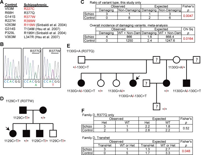
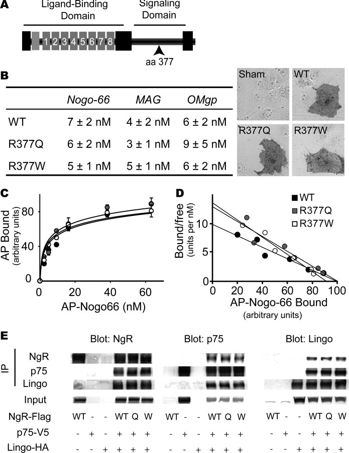
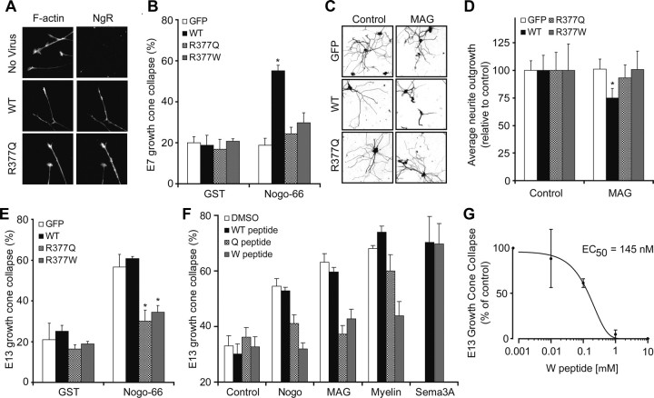
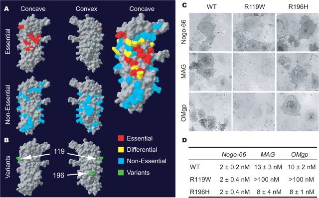
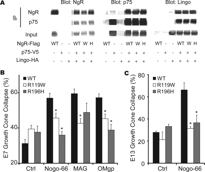
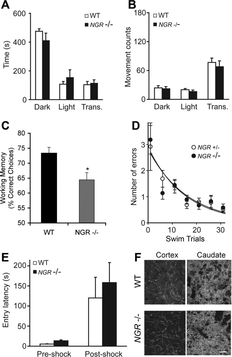
In schizophrenia, genetic predisposition has been linked to chromosome 22q11 and myelin-specific genes are misexpressed in schizophrenia. Nogo-66 receptor 1 (NGR or RTN4R) has been considered to be a 22q11 candidate gene for schizophrenia susceptibility because it encodes an axonal protein that mediates myelin inhibition of axonal sprouting. Confirming previous studies, we found that variation at the NGR locus is associated with schizophrenia in a Caucasian case-control analysis, and this association is not attributed to population stratification. Within a limited set of schizophrenia-derived DNA samples, we identified several rare NGR nonconservative coding sequence variants. Neuronal cultures demonstrate that four different schizophrenia-derived NgR1 variants fail to transduce myelin signals into axon inhibition, and function as dominant negatives to disrupt endogenous NgR1. This provides the first evidence that certain disease-derived human NgR1 variants are dysfunctional proteins in vitro. Mice lacking NgR1 protein exhibit reduced working memory function, consistent with a potential endophenotype of schizophrenia. For a restricted subset of individuals diagnosed with schizophrenia, the expression of dysfunctional NGR variants may contribute to increased disease risk.


Figures

Figure 1.
Figure 1.
NGR locus is associated with schizophrenia in a Caucasian case–control study. A, Linkage disequilibrium plot. The relative position of the five SNPs relative to the NGR locus are depicted. B, Association of specific single markers with the disease. The five markers selected were in Hardy–Weinberg equilibrium (p > 0.05). HWE, Hardy–Weinberg p value; MA, minor allele; control, allele frequency in the control samples; case, allele frequency in the schizophrenic samples. C, Association of the haplotype obtained by analyzing the five SNPs with the disease. For each haplotype, 0 represents the major allele and 1 represents the minor allele. The order of the SNPs in haplotypes does not follow the order of the chromosomal position; it follows this order: rs9606296, rs701421, rs701428, rs1567871, rs696880. Uncorrected p values and values corrected for multiple testing are listed.
Figure 2.
Figure 2.
NGR locus and genetic variation in schizophrenia. A, The NGR coding sequence from 542 schizophrenic individuals and 650 healthy controls was determined. The nonsynonymous coding sequence variants listed were present heterozygously in 12 individuals. Those variants listed in red are predicted to be “probably damaging” by the PolyPhen program. In addition, the four nonsynonymous coding sequence variants from two previous studies containing 328 schizophrenic individuals and 600 healthy controls are cited. B, 5′–3′ DNA sequence traces from a control individual with two WT NGR alleles, an affected individual heterozygous for a G>A transition at nucleotide 1130 resulting in the R377Q substitution, or an affected individual heterozygous for a C>T transition at nucleotide 1129 resulting in the R377W substitution. C, The observed numbers of coding variants predicted to be “probably damaging” or not by PolyPhen (http://genetics.bwh.harvard.edu/pph/) (Sunyaev et al., 2001) in the two groups is tabulated and analyzed statistically for the existing study. The metaanalysis table includes the individuals from previous studies and assesses the overall incidence of “probably damaging” variants in the two populations. D, E, Pedigree for R377W and R377Q families. Schizophrenic probands are indicated by the arrows. The filled symbols represent individuals with major psychiatric disease: schizophrenia, major depression with psychotic features, mild mental retardation with schizoid personality, or manic-depressive illness. F, The occurrence of R377Q variant or the transheterozygous R377Q variant in the presence of the 5′ noncoding change is tabulated for the family in D.
Figure 3.
Figure 3.
Schizophrenia-derived NgR1 signaling domain variants bind myelin ligands and interact with known coreceptors. A, Schematic structure of the NgR1 protein. The ligand-binding domain is composed of eight leucine-rich repeats. The segment from amino acid 311 to 450 is required for signaling through coreceptors and is anchored to the membrane via a GPI (glycosylphosphatidylinositol) moiety (7, 35). The site of amino acid 377 in the signaling domain is indicated. B, COS-7 cells were transfected with control vector, WT-NgR-expressing expression vector, R377Q-NgR1 vector, or R377W-NgR1 vector. The Kd for the binding of Nogo-66, MAG, and OMgp is reported. The binding of 10 nm AP-Nogo-66 ligand is detected as a dark reaction product on NgR1-expressing cells (right). C, AP-Nogo-66 binding assays were conducted over a range of ligand concentrations, and bound AP was measured. Error bars indicate SEM. D, The data from B are replotted. E, HEK293T cells were transfected with the indicated expression vectors (WT-NgR, WT; R377Q-NgR, Q; R377W-NgR, W). Lysates were immunoblotted directly or subjected to immunoprecipitation with the indicated antibodies and then immunoblotted. NgR1 amino acid variants do not significantly affect interaction with p75 or Lingo-1.
Figure 4.
Figure 4.
The R377Q and R377W-NgR1 variants are dominant-negative disruptors of myelin signaling. A, Chick E7 retinal neurons were cultured with or without herpes simplex virus directing the expression of WT-NgR, R377Q-NgR1, or R377W-NgR1 as indicated. The cultures were exposed to 100 nm GST (glutathione S-transferase)-Nogo-66, for 30 min, and then fixed and stained with phalloidin for F-actin and with anti-NgR1 antibody. Note that the viruses drive the expression of NgR1 protein. WT-NgR1 allows growth cones to collapse in response to Nogo-66, but R-377Q-NgR1 does not. B, The fraction of collapsed E7 retinal growth cones expressing WT-NgR1 is significantly greater than that for growth cones expressing GFP after exposure to 100 nm Nogo-66 (*p < 0.05, one-way ANOVA). C, Neurite outgrowth over 16 h from E20 rat DRG was assessed on a monolayer of control or MAG-expressing CHO cells after infection of neurons with virus expressing GFP, WT-NgR, R377Q-NgR1, or R377W-NgR. Neurons are visualized with anti-βIII-tubulin immunohistology. D, Mean neurite outgrowth from neurons expressing WT-NgR1 is significantly decreased on MAG cells relative to control monolayers (*p < 0.05, one-way ANOVA). Outgrowth from neurons expressing variant NgR1 is not reduced by MAG. E, The percentage of collapsed E13 chick DRG growth cones expressing the indicated proteins is reported from cultures similar to those in A. The values with R377Q-NgR1 and R377W-NgR1 virus are significantly different from those with control GFP virus (*p < 0.05, one-way ANOVA). F, Growth cone collapse of E13 chick DRG growth cones in the presence of 100 nm Nogo-66, 100 nm MAG-Fc, 3 μg/ml myelin protein, or 10 nm Sema3A. Collapse was assessed without peptide, with 1 μm WT peptide, with 1 μm R377Q peptide, or with 1 μm R377W peptide. Note that the R377W peptide suppresses collapse by Nogo-66, MAG, and myelin but not by Sema3A. Values with the variant NgR1 peptides are significantly different from those with the WT NgR1 peptide (*p < 0.05, one-way ANOVA). G, E13 chick DRG growth cone collapse in presence of 100 nm MAG-Fc and various concentrations of R377W peptide. All data are mean ± SEM from n = 3–7 experiments.
Figure 5.
Figure 5.
Ligand-binding domain in NgR1 and the effect of schizophrenia-derived sequence variants. A, The human NgR1 surface required for ligand binding is summarized from previous analysis of 74 Ala substitution variants (Park et al., 2006; Laurén et al., 2007). Residues required for Nogo-66, MAG, and OMgp are highlighted in red, and residues required for one ligand but not all are shown in yellow. B, Location of the two separate coding region variants identified in an Italian population (green) (Sinibaldi et al., 2004). The R119 residue is at the edge of the region required for ligand binding. The R196 residue is centered on the convex surface not implicated in ligand binding. C, Binding of AP-tagged myelin ligands to WT-NgR1, NgR-R119W, and NgR-R196H variants. Pictures shown are at the Kd for each ligand for WT-NgR. Note the absence of binding of AP-MAG and AP-OMgp to R119W-NgR. D, AP-Nogo-66, AP-MAG, and AP-OMgp binding assays were conducted over a range of ligand concentrations, and bound AP was measured.
Figure 6.
Figure 6.
NgR1 ligand-binding domain variants interact with known coreceptors but do not mediate myelin signaling. A, Interaction of NgR1 Italian variants with putative NgR1 coreceptors. HEK293T cells were transfected with the indicated expression vectors (WT-NgR, WT; R119W-NgR, W; R196H-NgR, H). Lysates were immunoblotted directly or subjected to immunoprecipitation and then immunoblot, as indicated. These NgR1 amino acid variations do not significantly alter interaction with p75 or Lingo-1. B, Growth cone collapse induced by myelin ligands in E7 retinal explants expressing WT-NgR1, R119W-NgR1, or R196H-NgR1. Values with variant NgR1-expressing virus are significantly different from those with wild-type NgR1 expression (*p < 0.05, one-way ANOVA). C, Dominant-negative effect of NgR1 variants in E13 cDRG expressing indicated constructs. Values with variant NgR1-expressing virus are significantly different from those with wild-type NgR1 expression (*p < 0.05, one-way ANOVA). All data are mean ± SEM from n = 3–6 experiments.
Figure 7.
Figure 7.
Selectively impaired working memory in NGR−/− mice. Mice were tested on a battery of tests to assess affective and cognitive responses. A, B, The light/dark exploration test was performed by placing the mouse in a cage (44 × 21 × 21 cm) that has one dark chamber (Dark) and one light chamber (Light). The animal was initially placed in the lighted side, and transitions between sides (trans) and the time spent in each chamber were recorded for 10 min using computer-monitored photocell beams. Mice were from a mixed 129 × C57BL/6 strain background. No significant difference of anxiety-like behavior was observed between WT and NGR−/− animals (n = 21, 10 WT, 11 −/−). C, The importance of NgR1 signaling to the spatial working memory functions of the prefrontal cortex in mice. NGR-deficient mice (−/−; n = 8) performed significantly worse on the spatial delayed alternation task than WT mice (+/+; n = 5). All mice were of a pure C57BL/6J genetic background (>10 backcrosses for the NGR mutation). Data represent mean ± SEM percentage correct over the 30 daily test sessions; *p = 0.017. D, NGR heterozygote and null littermate mice from a mixed strain background were subjected to a 2 d six-arm radial-arm water-maze paradigm as described previously (Morgan et al., 2000). To minimize odor cues, the goal arm was randomly assigned for each mouse. The start arm was varied for each trial, with the goal arm remaining constant. The mice were tested in the same manner on day 2. The number of incorrect arm entries (errors) was measured for 1 min. Mice made an arm choice within 20 s in every experiment. Each mouse's errors for five consecutive trials were averaged. E, Passive avoidance testing was performed in a mouse passive avoidance chamber (Ugo Basile). Mice used were from mixed 129 × C57BL/6 background. Testing occurred on 3 consecutive days; for all trials, the mouse was initially placed in the light chamber. On day 1, the mouse was allowed to move freely between the two compartments for 5 min. On day 2 (preshock), the entry latency into the dark chamber was measured; after entry, the door between the light and dark compartments closed, and a 2 s electric shock (0.2 mA) was administered through the grid floor. On day 3 (postshock), entry latency into the dark chamber was recorded. No differences in passive avoidance learning were observed in the WT and NGR−/− mice (n = 34, 17 WT, 17 −/−). F, Tyrosine hydroxylase-immunoreactive fibers in the prefrontal cerebral cortex and the caudate nucleus were visualized in sections from WT and NGR−/− adult mice. Scale bar, 100 μm. All data are mean ± SEM.
Figure 8.
Figure 8.
A small subset of mice lacking NgR1 exhibit decreased prepulse inhibition. Experiments used mice of a mixed background 129 × C57BL/6J with littermate controls (A–C) or a pure C57BL/6J genetic background (D–F). A, Acoustic startle response was measured as a function of stimulus intensity from mice of the indicated genotypes. B, Acoustic startle was measured with a stimulus of 110 dB stimulus preceded 100 ms by a pretone of the indicted sound level in NGR+/+ (n = 24), NGR+/− (n = 41), or NGR−/− (n = 38) mice. The trend to reduced NGR−/− PPI is not significant. C, Percentage of mice with absent PPI from the data set in B. Absent PPI is defined as <3% reduction in startle magnitude by prepulses of ≥85 dB. The incidence of absent PPI is greater in the NGR−/− population (*p < 0.05, χ2 test). D, Acoustic startle response was similar in NGR+/+ and NGR−/− mice (n = 57, 28 WT, and 29 NGR null). E, Prepulse inhibition of the acoustic startle was not significantly different in WT versus NGR−/− mice in this strain background, as opposed to the mixed background (n = 73, 34 WT, and 39 NGR null). F, The percentage of mice with absent PPI is reported. There is a significant increase in this percentage in the NGR−/− group (*p < 0.05, χ2). Error bars indicate SEM.

Comment in

References

    1. Akil M, Kolachana BS, Rothmond DA, Hyde TM, Weinberger DR, Kleinman JE. Catechol-O-methyltransferase genotype and dopamine regulation in the human brain. J Neurosci. 2003;23:2008–2013. - PMC - PubMed
    1. Baron M. Genetics of schizophrenia and the new millennium: progress and pitfalls. Am J Hum Genet. 2001;68:299–312. - PMC - PubMed
    1. Barton WA, Liu BP, Tzvetkova D, Jeffrey PD, Fournier AE, Sah D, Cate R, Strittmatter SM, Nikolov DB. Structure and axon outgrowth inhibitor binding of the Nogo-66 receptor and related proteins. EMBO J. 2003;22:3291–3302. - PMC - PubMed
    1. Bassett AS, Chow EW. 22q11 deletion syndrome: a genetic subtype of schizophrenia. Biol Psychiatry. 1999;46:882–891. - PMC - PubMed
    1. Bassett AS, Chow EW. Schizophrenia and 22q11.2 deletion syndrome. Curr Psychiatry Rep. 2008;10:148–157. - PMC - PubMed

Publication types

MeSH terms