. 2008 Dec 16;105(50):19915-9.
doi: 10.1073/pnas.0805803105. Epub 2008 Dec 3.

Carbonic anhydrase II-positive pancreatic cells are progenitors for both endocrine and exocrine pancreas after birth

Affiliations

Carbonic anhydrase II-positive pancreatic cells are progenitors for both endocrine and exocrine pancreas after birth

Akari Inada et al. Proc Natl Acad Sci U S A. .

Abstract

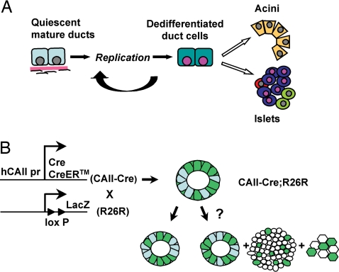
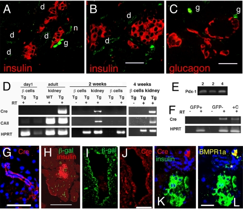
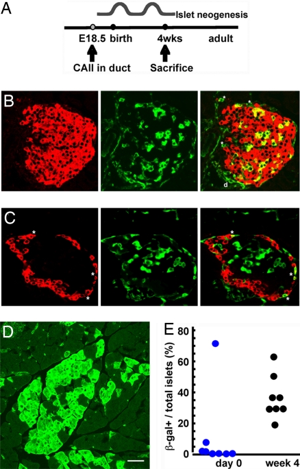
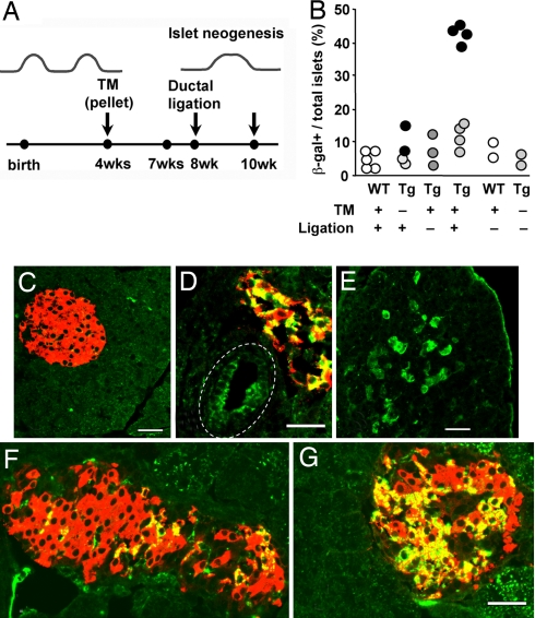
The regenerative process in the pancreas is of particular interest because diabetes results from an inadequate number of insulin-producing beta cells and pancreatic cancer may arise from the uncontrolled growth of progenitor/stem cells. Continued and substantial growth of islet tissue occurs after birth in rodents and humans, with additional compensatory growth in response to increased demand. In rodents there is clear evidence of pancreatic regeneration after some types of injury, with proliferation of preexisting differentiated cell types accounting for some replacement. Additionally, neogenesis or the budding of new islet cells from pancreatic ducts has been reported, but the existence and identity of a progenitor cell have been debated. We hypothesized that the progenitor cells are duct epithelial cells that after replication undergo a regression to a less differentiated state and then can form new endocrine and exocrine pancreas. To directly test whether ductal cells serve as pancreatic progenitors after birth and give rise to new islets, we generated transgenic mice expressing human carbonic anhydrase II (CAII) promoter: Cre recombinase (Cre) or inducible CreER(TM) to cross with ROSA26 loxP-Stop-loxP LacZ reporter mice. We show that CAII-expressing cells within the pancreas act as progenitors that give rise to both new islets and acini normally after birth and after injury (ductal ligation). This identification of a differentiated pancreatic cell type as an in vivo progenitor of all differentiated pancreatic cell types has implications for a potential expandable source for new islets for replenishment therapy for diabetes.


Conflict of interest statement

The authors declare no conflict of interest.

Figures

Fig. 1.
Fig. 1.
Experimental approach to test the hypothesis of ductal cells as pancreatic progenitors. (A) Our hypothesis is that with replication, mature duct cells regress to a less differentiated phenotype and then act as pancreatic progenitors to form new acini, islets, and ducts. (B) Tg mice in which human CAII promoter drives Cre or CreERTM expression were generated (CAII-Cre and CAII-CreERTM) and crossed with a Cre-mediated recombination reporter strain, R26R. In the double-Tg mice (CAII-Cre; R26R), if CAII-expressing ductal cells serve as pancreatic progenitors after birth, we should find β-galactosidase-positive cells not only in ducts but also in islets and acini; however, if duct cells do not act as progenitors, only ducts should be labeled.
Fig. 2.
Fig. 2.
Characterization of transgene expression. (A–C) At birth β-galactosidase immunostaining (green) was seen in ducts (d) and nerve ganglion (g) but not in insulin-positive beta cells (red; A and B), glucagon-positive cells (red; C) or acini of CAII-Cre mice. (D) GFP-expressing beta cells from CAII-Cre:MIP-GFP mice FACS purified for RNA analysis. At day 0 and at 2 and 4 weeks of age, the GFP+ sorted beta cells showed no band for Cre or CAII, even with 40 cycles of amplification. Kidneys from the transgenic (Tg) and non-Tg (WT) animals were positive and negative controls; RT − indicates controls for genomic contamination. (E) The 2- and 4-week RNA samples shown in D were probed for the transcription factor Pdx-1 at 28 cycles to show the integrity of RNA. (F) RNA from 10,000 pancreatic cells, either GFP+ beta cells or GFP cells from an 8-week-old CAII-Cre:MIP-GFP mouse, was similarly probed for Cre at 40 cycles. +C indicates positive control. (G) Cre immunostaining found in the cytoplasm and nuclei of small ducts (here 4 weeks) in CAII-Cre mice. (H) Expression of β-galactosidase in day 0 small ducts in CAII-Cre mice. (I and J) In CAII-CreERTM mice killed 3 weeks after the end of tamoxifen treatment, Cre and β-galactosidase immunostaining was found in larger ducts (I and J, adjacent sections). Cre staining here was cytoplasmic but not nuclear, and was only in some ductal cells. (K) The only other cells expressing Cre protein were ganglia (insulin, green; Cre, red). (L) The same section as shown in K with overlay of anti-BMPR1A also in green; ganglionic cells expressing BMPR1A are now yellow if expressing Cre or green (*) if not immunostained in previous image. (Scale bars, 50 μm in A–C, G, and I–L and 100 μm in H.)
Fig. 3.
Fig. 3.
Testing hypothesis of progenitors in normal neonatal development. (A) CAII starts being expressed at embryonic day 18.5. To test whether neogenesis occurs after birth, double-Tg mice were killed at 4 weeks of age after the expected waves of neogenesis (immediately after birth and around weaning). (B and C) At 4 weeks of age, some islets have costaining of β-galactosidase (green) and insulin (B), or glucagon (C) in red. Double-positive cells are shown as yellow in merged panels. (D) At 4 weeks of age, marked (β-galactosidase-positive) acinar cells are found as single scattered cells or in clusters localized to a lobe, suggesting new lobe formation since birth. (E) Quantification of positive islets/total islets, with each circle representing a single animal. At 4 weeks β-galactosidase+ insulin-positive cells were 16.7% ± 3.0% of all insulin-positive cells (mean: 345/2,268 insulin-positive cells per animal; 8 animals). (Scale bar, 50 μm.)
Fig. 4.
Fig. 4.
Testing hypothesis of progenitors in adult pancreatic regeneration in response to injury. (A) To determine whether new islets are formed in response to injury from duct cells of adult animals, the tamoxifen-inducible CAII-CreERTM double-Tg mice were implanted with 3-week tamoxifen pellets (TM) at 4 weeks of age, had washout period for about 1 week, had duct ligation at 8 weeks, and were killed 2 weeks later. (B) Quantification of positive islets/total islets, with each solid circle representing the mean of a single animal; for ligated Tg, values for pancreas distal to the ligation (dark circles) and for nonligated portion (light circles) are given. The last 2 columns are WT and double Tg at 6 months age, showing the stability of transgene without tamoxifen. In controls or the nonligated portion of the Tg with tamoxifen and ligation (C), there was little β-galactosidase (green) positivity, but distal to ligation in experimentals, some areas had β-galactosidase-marked ducts (D), acini (E), and islets (D, F, and G). In ligated portions, β-galactosidase+ insulin-positive cells were 23.6% ± 2.2% of all insulin-positive cells (mean: 673/2,997 insulin-positive cells; 4 animals), nonligated portions of the same animals were 5.5% ± 2.0% (mean: 110/821 insulin-positive), and in controls were 1.3% ± 1.2% (mean: 11/1,055 cells, 3 WT animals) and 0.9% ± 0.5% (mean: 17/1,785 cells, 4 Tg animals). (D) In this main duct (dashed line), Cre recombination occurred in about half the cells. (E) Expression of β-galactosidase was seen in acinar cells, often localized as to suggest new lobe formation. (F and G) The proportion of positive cells within the marked islets varied as seen in these islets. Shown are expression of β-galactosidase (green), insulin (red), and newly formed beta cells (yellow). (Scale bar, 50 μm.)

References

    1. Butler AE, et al. Beta-cell deficit and increased beta-cell apoptosis in humans with type 2 diabetes. Diabetes. 2003;52:102–110. - PubMed
    1. Li C, et al. Identification of pancreatic cancer stem cells. Cancer Res. 2007;67:1030–1037. - PubMed
    1. Bonner-Weir S, et al. The pancreatic ductal epithelium serves as a potential pool of progenitor cells. Pediatr Diabetes. 2005;5:15–22. - PubMed
    1. Wang RN, Kloppel G, Bouwens L. Duct- to islet-cell differentiation and islet growth in the pancreas of duct-ligated adult rats. Diabetologia. 1995;38:1405–1411. - PubMed
    1. Brockenbrough JS, Weir GC, Bonner-Weir S. Discordance of exocrine and endocrine growth after 90% pancreatectomy in rats. Diabetes. 1988;37:232–236. - PubMed

Publication types