. 2008 Dec 9;105(49):19199-204.
doi: 10.1073/pnas.0810634105. Epub 2008 Dec 3.

Enhancing nuclear receptor-induced transcription requires nuclear motor and LSD1-dependent gene networking in interchromatin granules

Affiliations

Enhancing nuclear receptor-induced transcription requires nuclear motor and LSD1-dependent gene networking in interchromatin granules

Qidong Hu et al. Proc Natl Acad Sci U S A. .

Erratum in

  • Proc Natl Acad Sci U S A. 2014 Feb 4;111(5):2046

Expression of concern in

Abstract

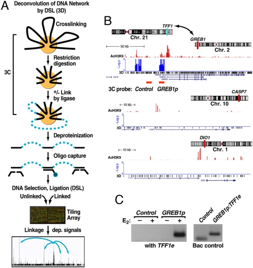
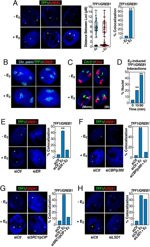
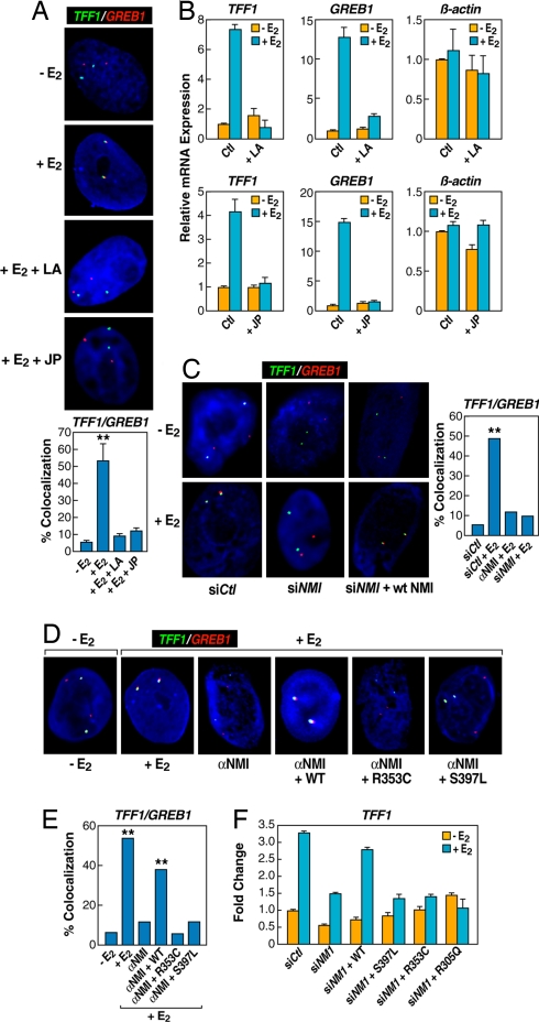
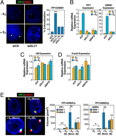
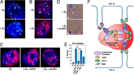
Although the role of liganded nuclear receptors in mediating coactivator/corepressor exchange is well-established, little is known about the potential regulation of chromosomal organization in the 3-dimensional space of the nucleus in achieving integrated transcriptional responses to diverse signaling events. Here, we report that ligand induces rapid interchromosomal interactions among specific subsets of estrogen receptor alpha-bound transcription units, with a dramatic reorganization of nuclear territories, which depends on the actions of nuclear actin/myosin-I machinery and dynein light chain 1. The histone lysine demethylase, LSD1, is required for these ligand-induced interactive loci to associate with distinct interchromatin granules, long thought to serve as "storage" sites for the splicing machinery, some critical transcription elongation factors, and various chromatin remodeling complexes. We demonstrate that this 2-step nuclear rearrangement is essential for achieving enhanced, coordinated transcription of nuclear receptor target genes.


Conflict of interest statement

The authors declare no conflict of interest.

Figures

Fig. 1.
Fig. 1.
Identification of long-range, estrogen induced chromosomal interactions by 3D. (A) Diagram of the 3D technology. The initial steps are identical to the established 3C technology. A key extension is DNA capturing by using a specific biotinylated oligonucleotide followed by DNA selection and ligation to detect cocaptured DNA fragments in a high-throughput and unbiased fashion. Specific signals were identified based on relative enrichment of DNA fragments linked by ligase compared with those from the parallel minus ligase control under an extensive dilution condition. (B) Specific interchromosomal interactions predicted by 3D. Three of the 6 assessed chromosomal intervals are shown, plotting the location of AcH3K9, an activation mark (red), and signal enrichment in 3D assay (blue) at the GREB1 locus (chromosome 2). Two negative controls (CASP7 in chromosome 10 and DIO1 in chromosome 1) show significant levels of AcH3K9 but no 3D signal. (C) 3C validation of the detected interchromosomal interaction predicted by 3D between TFF1 enhancer (chromosome 21) and GREB1 promoters (chromosome 2) in mock-treated and E2-induced (60 min) HMECs in which only the sample treated with E2 shows a strong interaction. (Right) Shows the primer efficiency obtained on randomly ligated BAC controls. The location of the 3C primers for GREB1 is indicted below the 3D signal track in B.
Fig. 2.
Fig. 2.
Rapid induction of interchromosomal interactions by nuclear hormone signaling. (A) 3D-FISH confirmation of E2-induced (60 min) TFF1:GREB1 interchromosomal interactions in HMECs with the distribution of loci distances measured (box plot with scatter plot) and quantification of colocalization (bar graph) before and after E2 treatment. Cells exhibiting mono- or biallelic interactions were combined for comparison with cells showing no colocalization; statistical significance in the bar graph was determined by χ2 test (**, P < 0.001). (B) 2D FISH confirmation of the interchromosomal interactions in MCF7 cells by combining chromosome paint (aqua) and specific DNA probes (green and red). (Upper) Illustrates two examples of mock-treated cells. (Lower) Shows the biallelic interactions/nuclear reorganization after E2 treatment for 60 min, exhibiting kissing events between chromosome 21 and chromosome 2. (C) Similar analysis on HMECs, but in this case using 3D FISH to paint chromosome 2 (red) and chromosome 21 (green), showing E2-induced chromosome 2–chromosome 21 interaction. Both assays revealed neither chromosome 21–chromosome 21 nor chromosome 2–chromosome 2 interactions in response to E2. (D) Temporal kinetics of GREB1:TFF1 interactions by 3D FISH in HMECs (**, P < 0.001 by χ2). (E–G) Nuclear microinjection of siRNA against ERα, CBP/p300, or SRC1/pCIP prevented E2-induced interchromosomal interactions, counting both mono- and biallelic interactions (**, P < 0.001 by χ2). The injection of siER and siDLC1 were done in the same experiment, sharing the same control group. (H) Nuclear microinjection of siRNA against LSD1, which was shown to be required for estrogen-induced gene expression (22), did not block E2-induced interchromosomal interactions. The injection of siLSD1 and SRC1/pCIP were done in a single experiment, sharing the same control group.
Fig. 3.
Fig. 3.
The nuclear actin/myosin machinery is required for long-distance chromosomal interactions. (A) Chemical disruption of actin polymerization with latrunculin (LA) (10 μM, 2 h), and prevention of actin depolymerization by jasplakinolide (JP) (10 μM, 2 h) impaired E2-induced interchromosomal interactions. The bar graph shows the percentage of cells ± SEM that showed colocalization under individual conditions (**, P < 0.001 by t test). (B) qPCR analysis of gene expression affected by JP and LA treatment. (C) Nuclear microinjection of siRNA or antibody against nuclear myosin I (NMI) abolished E2-induced TFF1:GREB1 interchromosomal interactions (**, P < 0.001 by χ2). (D and E) The requirement for the actin binding and ATPase activity of nuclear myosin I in mediating E2-induced interchromosomal interactions. Cells injected with antibody against NMI were coinjected with the plasmid expressing either WT or mutant NMI containing specific mutations in the myosin “head,” which were shown to be critical for actin binding (R353C) and ATPase activity (S497L) of the motor protein (**, P < 0.001 by χ2). (F) Rescue of E2-induced expression of TFF1 by expression of WT, but not mutant, NMI. Results are the average of triplicates ± SD differing by <10%; similar results were observed in duplicate experiments.
Fig. 4.
Fig. 4.
Enhanced gene expression resulting from interchromosomal interactions and the requirement for a key nuclear motor component. (A) Effects of nuclear microinjection of siRNA or antibody against DLCI on inhibiting E2-induced TFF1:GREB1 interactions (**, P < 0.001 by χ2). (B–D) Cells were treated with siRNAs against DLCI and/or BAF53, and the expression of specific genes as indicated, were quantified by RT-qPCR. Mean ± SD of triplicate determinations. (E) RNA FISH demonstrates the requirement for interchromosomal interactions to achieve enhanced, E2-induced gene expression. Quantification of expression from noninteracting (NI) and interacting allelic regions was based on the diameter of individual signals, which was then converted to volume (M ± SEM). Plot shows the significant increase of expression even in the absence of colocalization of either allele; and the comparison of interacting: noninteracting alleles (*, P < 0.01; **, P < 0.001 by t test).
Fig. 5.
Fig. 5.
Interchromatin granules are hubs for interchromosomal interactions. (A) 2D-FISH shows selective association of interacting loci with interchromatin granules. Only the interacting (TFF1:GREB1) alleles intermingle within interchromatin granules (ICGs) stained with αSC35 (pseudocolored blue), whereas the remaining noncolocalized alleles show no colocalization with ICGs. (B) 2D-FISH of biallelic TFF1/GREB1 interactions (purple/green as indicated by arrows) coincident with the IGCs. (C) LSD1 is required for the association of the interacting TFF1:GREB1 loci with IGCs (pseudocolored red). Microinjection of siRNA against LSD1 abolished the colocalization between the interacting TFF1:GREB1 loci and ICGs. (D) 3D-FISH demonstrating the requirement of LSD1 for association of the interacting TFF1:GREB1 loci (arrows) with IGCs. (E) Percentage of cells exhibiting IGC association in response to control and specific siRNA against LSD1 (**, P < 0.001 by χ2). The rescure experiments indicate that the enzymatic activity of LSD1 is at least partially required for mediating the association of the interacting gene loci with ICGs. (F) Proposed model of E2-induced, actin/myosin1/DLC1-mediated chromosomal movement and LSD1-dependent interactions with interchromatin granules, creating a 3-dimensional enhancer hub in the nucleus.

References

    1. Dennis AP, O'Malley BW. Rush hour at the promoter: How the ubiquitin proteasome pathway polices the traffic flow of nuclear receptor-dependent transcription. J Steroid Biochem Mol Biol. 2005;93:139–151. - PubMed
    1. Rosenfeld MG, Lunyak VV, Glass CK. Sensors and signals: A coactivator/corepressor/epigenetic code for integrating signal-dependent programs of transcriptional response. Genes Dev. 2006;20:1405–1428. - PubMed
    1. Carroll JS, et al. Chromosome-wide mapping of estrogen receptor binding reveals long-range regulation requiring the forkhead protein FoxA1. Cell. 2005;122:33–43. - PubMed
    1. Carroll JS, et al. Genome-wide analysis of estrogen receptor binding sites. Nat Genet. 2006;38:1289–1297. - PubMed
    1. Garcia-Bassets I, et al. Histone methylation dependent mechanisms impose ligand dependency for gene activation by nuclear receptors. Cell. 2007;128:505–518. - PMC - PubMed

Publication types

MeSH terms