Review
. 2008 Dec;72(4):590-641, Table of Contents.
doi: 10.1128/MMBR.00016-08.

ATP synthase and the actions of inhibitors utilized to study its roles in human health, disease, and other scientific areas

Affiliations
Review

ATP synthase and the actions of inhibitors utilized to study its roles in human health, disease, and other scientific areas

Sangjin Hong et al. Microbiol Mol Biol Rev. 2008 Dec.

Abstract

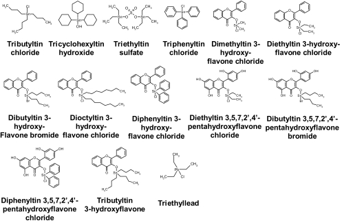
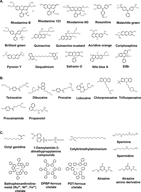
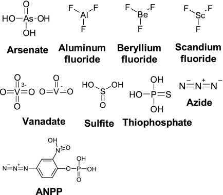
ATP synthase, a double-motor enzyme, plays various roles in the cell, participating not only in ATP synthesis but in ATP hydrolysis-dependent processes and in the regulation of a proton gradient across some membrane-dependent systems. Recent studies of ATP synthase as a potential molecular target for the treatment of some human diseases have displayed promising results, and this enzyme is now emerging as an attractive molecular target for the development of new therapies for a variety of diseases. Significantly, ATP synthase, because of its complex structure, is inhibited by a number of different inhibitors and provides diverse possibilities in the development of new ATP synthase-directed agents. In this review, we classify over 250 natural and synthetic inhibitors of ATP synthase reported to date and present their inhibitory sites and their known or proposed modes of action. The rich source of ATP synthase inhibitors and their known or purported sites of action presented in this review should provide valuable insights into their applications as potential scaffolds for new therapeutics for human and animal diseases as well as for the discovery of new pesticides and herbicides to help protect the world's food supply. Finally, as ATP synthase is now known to consist of two unique nanomotors involved in making ATP from ADP and P(i), the information provided in this review may greatly assist those investigators entering the emerging field of nanotechnology.


Figures

FIG. 1.
FIG. 1.
Current view of the structure of mitochondrial ATP synthase from metazoans. F1 is composed of α, β, γ, δ, and ɛ subunits, and F0 consists of a, b, c, d, e, f, g, A6L, and OSCP. IF1 is a regulatory protein. The coordinates of the subunits used in the structural model are 1E79 for the α, β, γ, δ, and ɛ subunits; 1ABV for the N-terminal domain of OSCP; 2CLY for F6, d, and the hydrophilic part of the b subunit; 1GMJ for IF1; and 1B9U for the transmembrane part of the b subunit. The ac10 subcomplex was modeled using the coordinates of the a and c subunits from 1C17, and the other subunits in the model were constructed manually using Quanta. No positions are assigned to the factor B and the e subunit. Here and where indicated in the other figure legends, the coordinates of protein structures were obtained from the PDB.
FIG. 2.
FIG. 2.
Structures of peptide inhibitors. (A) α-Helical basic peptide inhibitors. The coordinates of the inhibitors are 1BSN for the bacterial/chloroplast ɛ subunit, 1GMJ for IF1, and 2MLT for melittin. (B) Angiostatin and enterostatin. The coordinate for the structure is 1KI0. (C) Tentoxin and tentoxin analogs. (D) Leucinostatins and efrapeptins.
FIG. 3.
FIG. 3.
Structures of polyphenolic phytochemicals, estrogens, and structurally related compounds. (A) Stilbenes. SITS, 4-Acetamido-4′-isothiocyanostilbene 2,2′-disulfonate; DIDS, 4,4′-diisothiocyanatostilbene-2,2′-disulfonic acid. (B) Flavones and isoflavones. (C) Other polyphenolic phytochemicals. ECG, epicatechin gallate; EGCG, epigallocatechin gallate. (D) Steroidal estradiols and estrogen metabolites.
FIG. 4.
FIG. 4.
Structures of polyketide inhibitors.
FIG. 5.
FIG. 5.
Structures of organotin compounds and structural relatives.
FIG. 6.
FIG. 6.
Structures of polyenic α-pyrone derivatives.
FIG. 7.
FIG. 7.
Structures of cationic inhibitors. (A) Amphiphilic cationic dyes. EtBr, ethidium bromide. (B) TALAs and related compounds. (C) Other organic cations. DPBP, 4,4-diphenyl-2,2-bipyridine; PDT, 3-(2-pyridyl)-5,6-diphenyl-1,2,4-triazine.
FIG. 8.
FIG. 8.
Structures of phosphate analogs.
FIG. 9.
FIG. 9.
Structures of purine nucleotides and nucleotide analogs. (A) Nucleotides and nucleotide analogs. (B) Azidonucleotides.
FIG. 9.
FIG. 9.
Structures of purine nucleotides and nucleotide analogs. (A) Nucleotides and nucleotide analogs. (B) Azidonucleotides.
FIG. 9.
FIG. 9.
Structures of purine nucleotides and nucleotide analogs. (A) Nucleotides and nucleotide analogs. (B) Azidonucleotides.
FIG. 10.
FIG. 10.
Structures of amino acid residue modifiers. (A) Amino group modifiers. FDNB, 1-fluoro-2,4-dinitrobenzene. (B) Carboxyl group modifiers. CMCD, 1-cyclohexyl-3-(2-morpholinoethyl)carbodiimide metho-p-toluenesulfonate. (C) Cys/Tyr residue modifiers. DTNB, 5,5′-dithiobis(2-nitrobenzoic acid). (D) His residue modifiers.
FIG. 11.
FIG. 11.
Miscellaneous inhibitors. TCS, tetrachlorosalicylanilide.
FIG. 12.
FIG. 12.
Inhibitory sites of ATP synthase. The inhibitor binding sites in the ATP synthase as revealed by biochemical/structural studies are indicated by red circles, and the binding subunits in which the binding sites have not been completely clarified are indicated by green circles. The coordinates of each subunit in the structural model are the same as in Fig. 1.

References

    1. Abad, M. C., R. K. Arni, D. K. Grella, F. J. Castellino, A. Tulinsky, and J. H. Geiger. 2002. The X-ray crystallographic structure of the angiogenesis inhibitor angiostatin. J. Mol. Biol. 3181009-1017. - PubMed
    1. Abrahams, J. P., S. K. Buchanan, M. J. Van Raaij, I. M. Fearnley, A. G. Leslie, and J. E. Walker. 1996. The structure of bovine F1-ATPase complexed with the peptide antibiotic efrapeptin. Proc. Natl. Acad. Sci. USA 939420-9424. - PMC - PubMed
    1. Ackerman, S. H., C. Grubmeyer, and P. S. Coleman. 1987. Evidence for catalytic cooperativity during ATP hydrolysis by beef heart F1-ATPase. Kinetics and binding studies with the photoaffinity label BzATP. J. Biol. Chem. 26213765-13772. - PubMed
    1. Agarwal, N., and V. K. Kalra. 1984. Studies on the mechanism of action of local anesthetics on proton translocating ATPase from Mycobacterium phlei. Biochim. Biophys. Acta 764316-323. - PubMed
    1. Ahmad, Z., and A. E. Senior. 2006. Inhibition of the ATPase activity of Escherichia coli ATP synthase by magnesium fluoride. FEBS Lett. 580517-520. - PubMed

Publication types