. 2008;3(12):e3842.
doi: 10.1371/journal.pone.0003842. Epub 2008 Dec 4.

CTLA-4 activation of phosphatidylinositol 3-kinase (PI 3-K) and protein kinase B (PKB/AKT) sustains T-cell anergy without cell death

Affiliations

CTLA-4 activation of phosphatidylinositol 3-kinase (PI 3-K) and protein kinase B (PKB/AKT) sustains T-cell anergy without cell death

Helga Schneider et al. PLoS One. 2008.

Erratum in

  • PLoS ONE. 2009;4(1). doi: 10.1371/annotation/5f36e0a6-8e8b-4b33-8e96-a2d53d5c1e46

Abstract

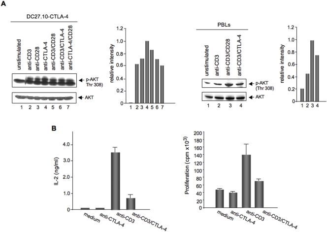
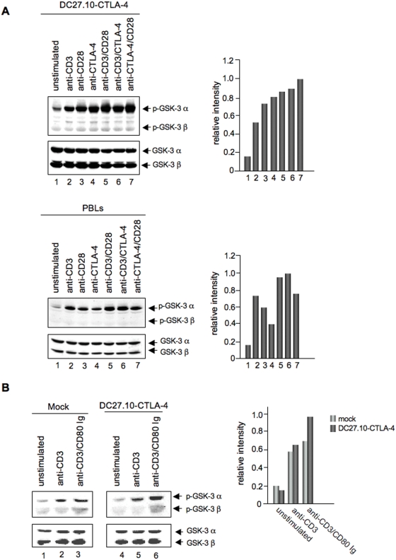
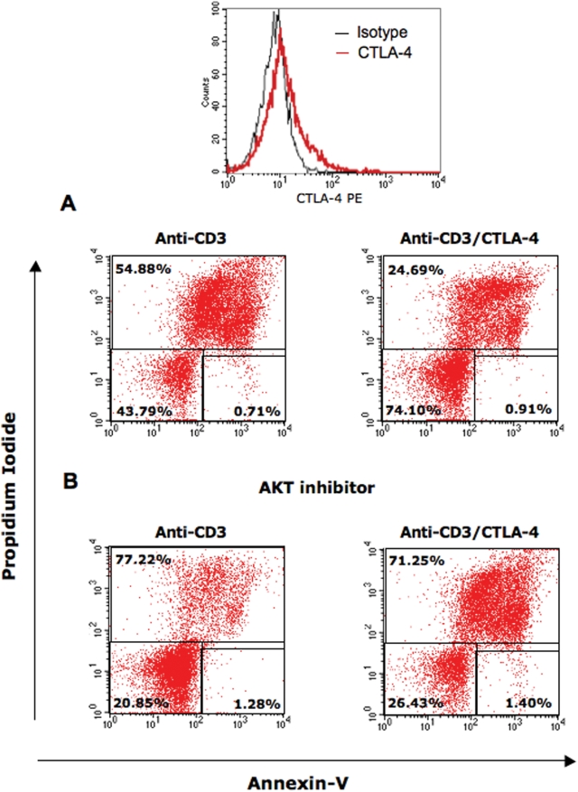
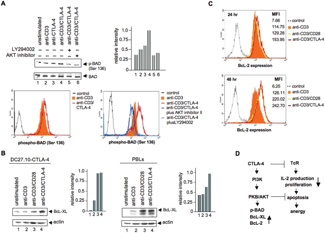
The balance of T-cell proliferation, anergy and apoptosis is central to immune function. In this regard, co-receptor CTLA-4 is needed for the induction of anergy and tolerance. One central question concerns the mechanism by which CTLA-4 can induce T-cell non-responsiveness without a concurrent induction of antigen induced cell death (AICD). In this study, we show that CTLA-4 activation of the phosphatidylinositol 3-kinase (PI 3-K) and protein kinase B (PKB/AKT) sustains T-cell anergy without cell death. CTLA-4 ligation induced PI 3K activation as evidenced by the phosphorylation of PKB/AKT that in turn inactivated GSK-3. The level of activation was similar to that observed with CD28. CTLA-4 induced PI 3K and AKT activation also led to phosphorylation of the pro-apoptotic factor BAD as well as the up-regulation of BcL-XL. In keeping with this, CD3/CTLA-4 co-ligation prevented apoptosis under the same conditions where T-cell non-responsiveness was induced. This effect was PI 3K and PKB/AKT dependent since inhibition of these enzymes under conditions of anti-CD3/CTLA-4 co-ligation resulted in cell death. Our findings therefore define a mechanism by which CTLA-4 can induce anergy (and possibly peripheral tolerance) by preventing the induction of cell death.


Conflict of interest statement

Competing Interests: The authors have declared that no competing interests exist.

Figures

Figure 1
Figure 1
Panel A: CTLA-4 induces phosphorylation of PKB/AKT. Upper left panels: DC27.10-CTLA-4 were either left untreated (lane 1) or stimulated for 30 min with anti-CD3 (lane 2), anti-CD28 (lane 3), anti-CTLA-4 (lane 4), anti-CD3/CD28 (lane 5), anti-CD3/CTLA-4 (lane 6) and anti-CD28/CTLA-4 (lane 7) antibodies. Cell lysates were immunoblotted with anti-phospho-AKT (Thr-308) antibody (lanes 1–7). Histogram depiction of phosphorylated AKT as detected by densitometric reading. Lower panel: Equal amounts of cell lysates were immunoblotted for AKT (lanes 1–7). Upper right panels: Pre-activated T-cells were either left untreated (lane 1) or stimulated for 30 min with anti-CD3 (lane 2), anti-CD3/CD28 (lane 3) and anti-CD3/CTLA-4 (lane 4) antibodies. Cell lysates were immunoblotted with anti-phospho-AKT (Thr-308) (lanes 1–4) antibody. Histogram depiction of phosphorylated AKT as detected by densitometric reading. Lower panel: Equal amounts of cell lysates were immunoblotted for AKT (lanes 1–4). Panel B: CTLA-4 mediated inhibition of IL-2 production and proliferation. DC27.10-CTLA-4 cells and peripheral T-cells were either left unstimulated, or stimulated with anti-CTLA-4, anti-CD3 and anti-CD3/CTLA-4 mAbs. After 24 hours, IL-2 production was measured by ELISA. After 48 hours, proliferation was measured by [3H] thymidine incorporation. Bar graphs show mean±SD. Results are representative of at least three experiments.
Figure 2
Figure 2
Panel A: CTLA-4 mediated phosphorylation of GSK-3. Upper left panel: DC27.10-CTLA-4 and pre-activated peripheral T-cells were stimulated for 30 min as described for Figure 1A. Cell lysates were immunoblotted with anti-phospho-GSK-3 α/β antibody (lanes 1–7). Upper right panel: Histogram depiction of phosphorylated GSK-3 as detected by densitometric reading. Lower left panel: Cells treated as described above were lysed and immunoblotted with an antibody against total GSK-3 α/β (lanes 1–7). Similar results were obtained from at least three other experiments. Lower right panel: Histogram depiction of phosphorylated GSK-3 as detected by densitometric reading. Panel B: Ligation of CTLA-4 by natural ligand induces phosphorylation of GSK-3. DC27.10 cells transfected with mock (lanes 1–3) or CTLA-4 (lanes 4–6) were either left untreated (lanes 1, 4) or stimulated for 30 min with anti-CD3 (lanes 2, 5) or anti-CD3/CD80Ig (lanes 3, 6) and assesssed for phosphorylation of GSK-3 by immunoblotting with anti-phospho-GSK-3 α/β antibody (lanes 1–6). Lower panel: Equal amounts of cell lysates were immunoblotted for total GSK-3 α/β (lanes 1–6). Right panel: Histogram depiction of phosphorylated GSK-3 as detected by densitometric reading. Results are representative of at least two experiments.
Figure 3
Figure 3. CTLA-4 ligation rescues cells from apoptosis and is dependent on AKT activation.
Pre-activated PBLs were stimulated with anti-CD3 (left panel) and anti-CD3/CTLA-4 (right panel) in the absence (upper panel) or presence of AKT inhibitor (AKT inhibitor II) (lower panel). 48 hours later, cells were stained with Annexin V-Cy5 and PI and analysed by FACS for cell death. Top panel shows CTLA-4 surface expression in these cells. Similar results were obtained from at least three other experiments.
Figure 4
Figure 4
Panel A: CTLA-4 ligation induces phosphorylation of BAD at Ser-136. Upper panel: DC27.10-CTLA-4 cells were either left unstimulated (lane 1) or stimulated for 30 min with anti-CD3 (lane 2), anti-CTLA-4 (lane 3) and anti-CD3/CTLA-4 (lane 4) mAbs. In lane 5 and 6, cells were pretreated with LY 294002 (100 µM, 30 min) or AKT inhibitor II (15 µM, 30 min), respectively and then stimulated with anti-CD3/CTLA-4 antibodies. Cell lysates were immunoblotted with anti-phospho-BAD (Ser-136) antibody (lanes 1–6). Right upper panel: Histogram depiction of phospho-BAD as detected by densitometric reading. Middle panel: Similar amounts of cell lysates were immunoblotted for total BAD (lanes 1–6). Lower panel: Pre-activated PBLs were re-stimulated with anti-CD3 or anti-CD3/CTLA-4 in the absence or presence of AKT inhibitor II or LY 294002. 24 hours later, cells were washed, stained with anti-phospho-BAD (Ser 136)/anti-rabbit AlexaFluo488 antibodies and analysed by flow cytometry. Panel B: CTLA-4 ligation induces up-regulation of BcL-XL. Left panel: DC27.10-CTLA-4 cells were either left unstimulated (lane 1) or stimulated for 24 hours with anti-CD3 (lane 2), anti-CD3/CD28 (lane 3), and anti-CD3/CTLA-4 (lane 4) antibodies. Cell lysates were immunoblotted with anti-BcL-XL antibody (lanes 1–4). Right panel: Pre-activated peripheral T-cells were treated as described above and assessed for BcL-XL expression by immunoblotting with anti-BcL-XL antibody (lanes 1–4). Middle panels: Similar amounts of cell lysates were immunoblotted for actin (lanes 1–4). Panel C: CTLA-4 ligation induces up-regulation of BcL-2. Pre-activated PBLs were re-stimulated with anti-CD3, anti-CD3/CD28 or anti-CD3/CTLA-4 antibodies. 24 and 48 hours later, cells were washed, stained with anti-BcL-2/anti-rabbit AlexaFluo647 antibodies and analysed by flow cytometry. Similar results were obtained from three other experiments. Panel D: CTLA-4 induced pro-survival signaling pathways. CTLA-4 can increase cell survivial under conditions of anti-CD3/CTLA-4 induced non-responsiveness. CTLA-4-PI 3K activates PKB/AKT by phosphorylation at Thr-308 that in turn inactivates pro-apoptotic BAD by phosphorylation at Ser-136. Inhibitors of PI 3K and PKB/AKT blocked this event. Decreased active BAD induced by CTLA-4 ligation was accompanied by increased levels of BcL-XL/BcL-2 expression. BcL-XL/BcL-2 are then able to mediate their mitochondrial-dependent pro-survival effects.

References

    1. Linsley PS. Distinct roles for CD28 and cytotoxic T lymphocyte-associated molecule 4 receptor during T-cell activation? J. Exp. Med. 1995;182:289–292. - PMC - PubMed
    1. Thompson CB, Allison JP. The emerging role of CTLA-4 as an immune attenuator. Immunity. 1997;7:445–450. - PubMed
    1. Rudd CE, Schneider H. Unifying concepts in CD28, ICOS and CTLA-4 co-receptor signalling. Nat. Rev. Immunol. 2003;3:544–556. - PubMed
    1. Linsley PS, Greene JL, Brady W, Bajorath J, Ledbetter JA, et al. Human B7-1 (CD80) and B7-2 (CD86) bind with similar avidities but distinct kinetics to CD28 and CTLA-4 receptors. Immunity. 1994;1:793–801. - PubMed
    1. Krummel MF, Allison JP. CD28 and CTLA-4 have opposing effects on the response of T cells to stimulation. J. Exp. Med. 1995;182:459–465. - PMC - PubMed

Publication types