. 2008:14:2190-203.
Epub 2008 Dec 3.

Neuroprotective effects of cannabidiol in endotoxin-induced uveitis: critical role of p38 MAPK activation

Affiliations

Neuroprotective effects of cannabidiol in endotoxin-induced uveitis: critical role of p38 MAPK activation

A B El-Remessy et al. Mol Vis. 2008.

Abstract

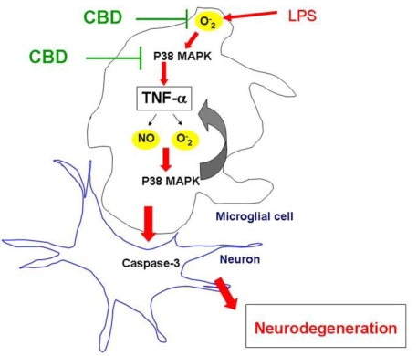
Purpose: Degenerative retinal diseases are characterized by inflammation and microglial activation. The nonpsychoactive cannabinoid, cannabidiol (CBD), is an anti-inflammatory in models of diabetes and glaucoma. However, the cellular and molecular mechanisms are largely unknown. We tested the hypothesis that retinal inflammation and microglia activation are initiated and sustained by oxidative stress and p38 mitogen-activated protein kinase (MAPK) activation, and that CBD reduces inflammation by blocking these processes.

Methods: Microglial cells were isolated from retinas of newborn rats. Tumor necrosis factor (TNF)-alpha levels were estimated with ELISA. Nitric oxide (NO) was determined with a NO analyzer. Superoxide anion levels were determined by the chemiluminescence of luminol derivative. Reactive oxygen species (ROS) was estimated by measuring the cellular oxidation products of 2', 7'-dichlorofluorescin diacetate.

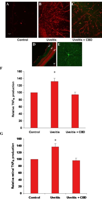
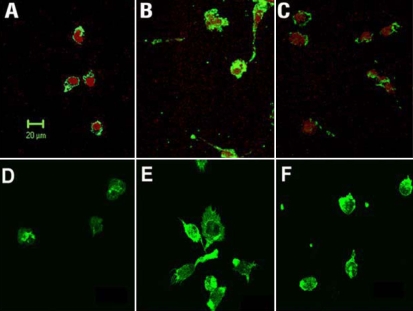
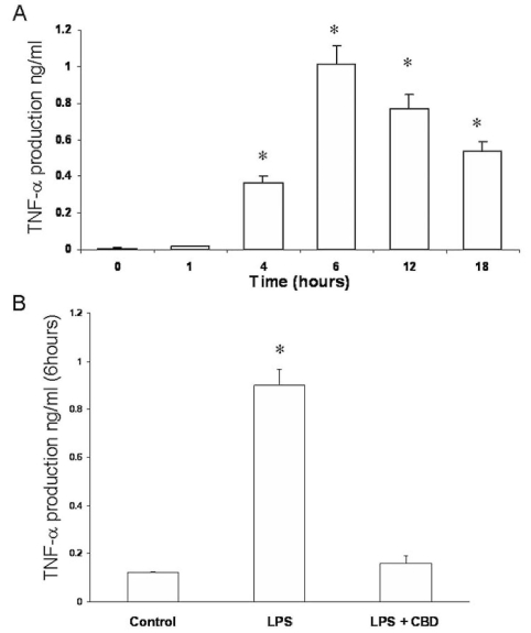
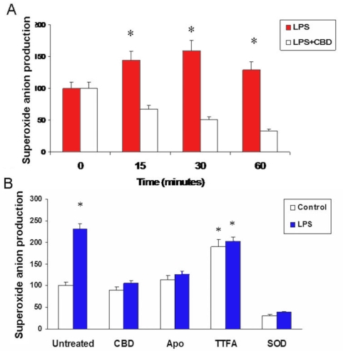
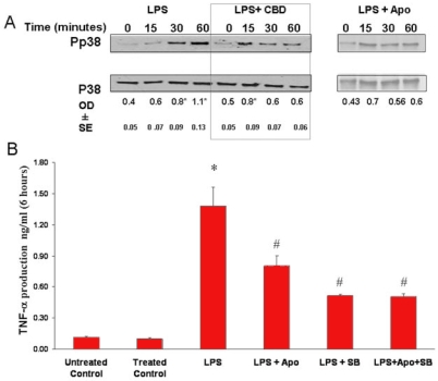
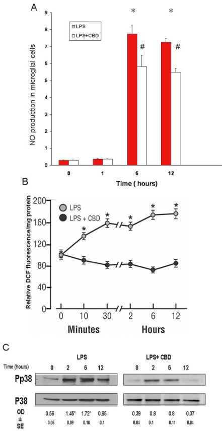
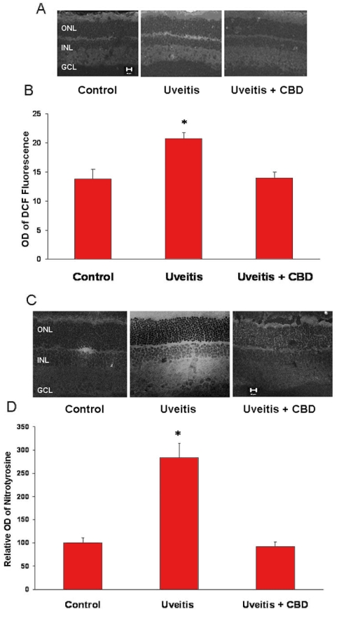
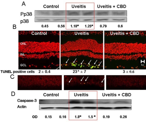
Results: In retinal microglial cells, treatment with lipopolysaccharide (LPS) induced immediate NADPH oxidase-generated ROS. This was followed by p38 MAPK activation and resulted in a time-dependent increase in TNF-alpha production. At a later phase, LPS induced NO, ROS, and p38 MAPK activation that peaked at 2-6 h and was accompanied by morphological change of microglia. Treatment with 1 microM CBD inhibited ROS formation and p38 MAPK activation, NO and TNF-alpha formation, and maintained cell morphology. In addition, LPS-treated rat retinas showed an accumulation of macrophages and activated microglia, significant levels of ROS and nitrotyrosine, activation of p38 MAPK, and neuronal apoptosis. These effects were blocked by treatment with 5 mg/kg CBD.

Conclusions: Retinal inflammation and degeneration in uveitis are caused by oxidative stress. CBD exerts anti-inflammatory and neuroprotective effects by a mechanism that involves blocking oxidative stress and activation of p38 MAPK and microglia.


Figures

Figure 1
Figure 1
Cannabidiol prevents retinal microglial activation or macrophage infiltration and inhibits serum and retinal tumor necrosis factor release in the uveitic rat. A-E: Confocal micrographs of retina whole-mounts or sections that show activated microglia or infiltrated macrophages as stained by the microglia/macrophage-specific marker CD11b and by Texas red- or Oregon green-conjugated secondary antibody. A: Normal rat. B: Twenty-four h after lipopolysaccharide (LPS) injection. C: Cannabidiol (CBD)-pretreated and LPS-injected. D: Microglia in normal eye sections, Oregon green, counter-stained with propidium iodide (red). E: Microglia in normal retinal whole-mounts, Oregon green. F: Serum TNF-α levels in 3 rats, 24 h after LPS injection with or without CBD treatment (mean±SEM; asterisk represents that it is significantly different when compared with the control at p<0.05). G: Retinal TNF-α levels in 3 rats, 24 h after LPS injection with or without CBD treatment (mean±SEM; asterisk represents that it is significantly different when compared with the control at p<0.05).
Figure 2
Figure 2
Effects of cannabidiol (CBD) on lipopolysaccharide (LPS)-induced morphological changes in rat retinal microglial cells. Confocal micrographs were made of cells identified by microglial- and macrophage-specific marker CD11b followed by Oregon green-conjugated secondary antibody. Cell nuclei were counter-stained by propidium iodide. A: Cells were cultured in serum-free medium for 12 h. B: Cells were treated with 30 ng/ml lipopolysaccharide in serum-free medium for 12 h. C: Cells were cannabidiol (CBD, 1 μM)-pretreated and lipopolysaccharide-treated for 12 h. D-F: Cells similarly treated were also stained with fluorescent phallotoxins for Filamentous actin distribution and cellular morphology. Scale bar represents 20 μm.
Figure 3
Figure 3
Cannabidiol reduces tumor necrosis factor-α levels in lipopolysaccharide-treated rat retinal microglial cells. Culture medium of retinal microglial cells were collected at different time points after lipopolysaccharide (LPS) treatment and assayed for tumor necrosis factor-α (TNF-α) with ELISA. A: TNF-α levels were measured at different times in LPS-treated microglial cells and were compared with the control. B: TNF-α levels were compared in the presence or absence of 1 μM cannabidiol (CBD). Data shown is the mean of 6 samples measured at 6 h±SEM. Asterisk represents that it is significantly different when compared with the 0 time or control at p<0.05.
Figure 4
Figure 4
Cannabidiol blocks lipopolysaccharide-induced early superoxide formation A: In microglial cells treated with lipopolysaccharides (LPS), maximal reactive oxygen species (ROS) formation measured by chemiluminescence assay was observed at 30 min. Cannabidiol (CBD) reduced ROS formation during this period. Data shown is the mean of 5 samples±SEM; Asterisk represents that it is significantly different when compared with CBD-treated at p<0.01. B: Comparison of ROS formation measured by chemiluminescence assay on microglial cells pretreated with CBD, apocynin, TTFA, or PEG-SOD and treated with vehicle (control) or LPS. Results suggested that superoxide component of ROS in LPS-treated retinal microglia is generated from NADPH oxidase. Data shown is the mean of 6 samples measured at 30 min±SEM; Asterisk represents that it is significantly different when compared with CBD-treated at p<0.01.
Figure 5
Figure 5
Oxidative stress and p38 MAPK activation are causally related and are involved in release of tumor necrosis factor-α. A: Lipopolysaccharide (LPS) caused a time-dependent activation of p38 MAPK with highest activation at 60 min. Cannabidiol (CBD; 1 μM) or apocynin (200 μM) reduced the activation of p38 MAPK (phospho-p38) throughout the 60 min. Data shown is the mean of 2-3 experiments±SEM; asterisk represents that it is significantly different when compared with the controls at p<0.05. B: Pretreatment of microglial cells with apocynin (200 μM) or the p38 MAPK inhibitor, SB203580 (10 µM), inhibited the LPS-induced release of tumor necrosis factor-α (TNF-α). Treatment with both apocynin and SB203580 did not further decrease the release of TNF-α. Data shown is the mean of 4-8 experiments±SEM. Asterisk represents that it is significantly different at p<0.001 when compared with vehicle control or with apocynin-treated and SB203580-treated control; hash mark represents that it is significantly different at p<0.05 when compared with LPS alone.
Figure 6
Figure 6
Cannabidiol reduces lipopolysaccharide-induced late increases of nitric oxide and peroxynitrite and p38 MAPK activation. A: Lipopolysaccharide (LPS) caused maximal increase in nitric oxide (NO) formation at 6 h and after. Cannabidiol (CBD) reduced NO formation during this period. Data shown is the mean of 4-6 experiments±SEM. Asterisk represents that it is significantly different at p<0.05 when compared with 0 time. B: LPS caused peroxynitrite formation as early as 15 min, followed by a further increase at 6-12 h. CBD reduced peroxynitrite formation during this period. Data shown is the mean of 6 experiments±SEM; asterisk represents that it is significantly different at p<0.005 as compared to CBD-treated. C: LPS treatment of microglial cells for 0-12 h induced a second peak of phospho-p38 MAPK at 2-6 h. CBD significantly reduced p38 MAPK activation level during this period. Data shown is the mean of 3 experiments±SEM; asterisk represents that it is significantly different at p<0.05 when compared with 0 time.
Figure 7
Figure 7
Cannabidiol reduces oxidative and nitrative stresses in the uveitic retina. A: Cannabidiol (CBD) reduces reactive oxygen species (ROS) in the retinas of uveitic rats as represented by DCF fluorescence in rat retina. Representative image shows the fluorescence distribution in different retinal layers (magnification ×100). Abbreviations: Ganglion cell layer (GCL); inner nuclear layer (INL); outer nuclear layer (ONL). B: Morphometric analysis of fluorescence intensity in serial sections of rat eyes shows that uveitic rats had a significant increase in fluorescence (1.5-fold) compared with controls. Treatment with CBD (5 mg/kg) inhibited ROS formation in uveitic rats. Data shown is the mean±SEM of 4-5 animals in each group (asterisk represents that it is significantly different when compared with the controls at p<0.05). C: CBD reduces nitrotyrosine in the retina of uveitic rats. A representative image shows the nitrotyrosine distribution mainly in the retinal plexiform layers and outer segments (magnification ×200). D: Morphometric analysis of fluorescence intensity in serial sections of rat eyes showing that uveitic rats had a significant increase in fluorescence (2.8-fold) compared with controls. Treatment with CBD (5 mg/kg) inhibited nitrotyrosine formation in the uveitic rats. Data shown is the mean±SEM of 6 animals in each group (asterisk represents that it is significantly different when compared with the controls at p<0.05).
Figure 8
Figure 8
Cannabidiol reduces p38 MAPK activation and prevents cell death in the uveitic retina. A: Lipopolysaccharide (LPS) treatment of rats resulted in a significant increase in p38 MAPK activation at 24 h. CBD (5 mg/kg) treatment reduced this effect. Data from 2 animals in each group are shown (asterisk represents that it is significantly different at p<0.05 as compared to control), Similar results were obtained in 2 independent experiments. B: CBD blocked neuronal cell death, as detected by TUNEL analysis. Data shown is the mean±SEM of 3 animals in each group (asterisk represents that it is significantly different at p<0.05 as compared to control). C: Colocalization of phospho-p38 MAPK (red) and TUNEL+cells (green) in the retinal ganglion cell layer. CBD blocked LPS-induced activation of p38 MAPK and blocked neuronal cell death. D: CBD blocked LPS-induced caspase-3 expression that was detected by Western analysis in the uveitic retina. Data from 2 animals in each group are shown (asterisk represents that it is significantly different at p<0.05 compared to control). Similar results were obtained in 2 independent experiments.
Figure 9
Figure 9
Schematic figure summarizes the proposed mechanism of lipopolysaccharide-induced retinal degeneration. Lipopolysaccharide (LPS)-induced oxidative stress leads to p38 MAPK activation, tumor necrosis factor-α (TNF-α) release, and another phase of p38 MAPK activation. The autoregulatory loop of TNF-α release and oxidative stress leads to microglial activation and retinal neurodegeneration. Suggested sites where cannabidiol (CBD) blocks this pathway are indicated.

References

    1. Langmann T. Microglia activation in retinal degeneration. J Leukoc Biol. 2007;81:1345–51. - PubMed
    1. Medana IM, Chan-Ling T, Hunt NH. Redistribution and degeneration of retinal astrocytes in experimental murine cerebral malaria: relationship to disruption of the blood-retinal barrier. Glia. 1996;16:51–64. - PubMed
    1. Hoekzema R, Verhagen C, van Haren M, Kijlstra A. Endotoxin-induced uveitis in the rat. The significance of intraocular interleukin-6. Invest Ophthalmol Vis Sci. 1992;33:532–9. - PubMed
    1. McMenamin PG, Crewe J. Endotoxin-induced uveitis. Kinetics and phenotype of the inflammatory cell infiltrate and the response of the resident tissue macrophages and dendritic cells in the iris and ciliary body. Invest Ophthalmol Vis Sci. 1995;36:1949–59. - PubMed
    1. Arai K, Wood JP, Osborne NN. Beta-adrenergic receptor agonists and antagonists counteract LPS-induced neuronal death in retinal cultures by different mechanisms. Brain Res. 2003;985:176–86. - PubMed

Publication types

MeSH terms

LinkOut - more resources