. 2009 Mar-Apr;15(3-4):76-84.
doi: 10.2119/molmed.2008.00108. Epub 2008 Nov 19.

CD38 and CD157 ectoenzymes mark cell subsets in the human corneal limbus

Affiliations

CD38 and CD157 ectoenzymes mark cell subsets in the human corneal limbus

Alberto L Horenstein et al. Mol Med. 2009 Mar-Apr.

Abstract

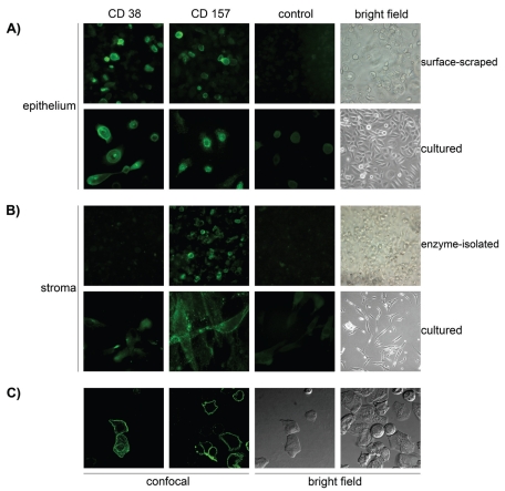
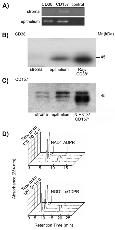
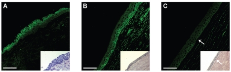
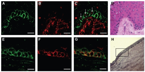
Nicotinamide adenine dinucleotide (NAD(+)), a precursor of molecules involved in cell regulatory processes, is released in extra-cellular compartments after stress or inflammation.This study investigates the expression in the human cornea of CD38 and CD157, two NAD(+)-consuming ectoenzymes and surface receptors. The analysis in corneal epithelial and stromal cells was performed by means of multiple approaches, which included immunofluorescence, reverse transcriptase polymerase chain reaction (RT-PCR), Western blot, and confocal microscopy. The presence of enzymatically active NAD(+)-consumers in intact corneal cells was analyzed by high performance liquid chromatography (HPLC)-based assays. The results obtained show that CD38 and CD157 are expressed constitutively by corneal cells: CD38 appears as a 45-kDa monomer, while CD157 is a 42- to 45-kDa doublet. The molecules are enzymatically active, with features reminiscent of those observed in human leukocytes. CD38 is expressed by cells of the suprabasal limbal epithelium, whereas it is not detectable in central corneal epithelium and stroma. CD157 is expressed by basal limbal clusters, a p63(+)/cytokeratin 19(+) cell subset reported to contain corneal stem cells, and by stromal cells. The results of the work indicates that the human cornea is equipped with molecular tools capable of consuming extracellular NAD(+), and that CD157 is a potential marker of corneal limbal cells in the stem cell niche. The presence and characteristics of these ectoenzymes may be exploited to design drugs for wound repair or for applications in tissue transplantation.


Figures

Figure 1
Figure 1
Immunodetection of the CD38 and CD157 molecules expressed by human corneal cells. Isolated human corneal cells were stained with anti-CD38 (AT13/5, IgG1), anti-CD157 (SY/11B5, IgG1) mAbs, or with an isotype-matched irrelevant control mAb. (A) Staining of CD38 and CD157 with specific mAbs in surface-scraped or cultured epithelial cells, with corresponding control and bright field images. (B) Staining of CD38 and CD157 in enzyme-isolated or cultured stromal cells, with corresponding control and bright field images. (C) Fluorescent images acquired using confocal microscopy, showing the staining of CD38 and CD157 in surface-scraped corneal epithelial cells and corresponding fields seen with interference contrast. The molecules are localized at the membrane levels of the corneal epithelial cells.
Figure 2
Figure 2
Biochemical analysis of the CD38 and CD157 molecules expressed by human corneal cells. (A) RT-PCR profile of CD38 and CD157 transcripts in corneal cells. PCR products were amplified from cDNA obtained from epithelial and stromal cells using CD38- or CD157-specific primer sets. Negative controls lacking cDNA template were used to rule out contamination. (B–C) Western blot analysis of corneal stromal and epithelial proteins immunoprecipitated with mAbs to CD38 and CD157. Precipitates of epithelial and stromal cells from biopsied corneas with anti-CD38 (IB4, IgG2a) or with anti-CD157 (SY/11B5, IgG1) mAbs were analyzed by 4% to 12% SDS-PAGE under non-reducing conditions and blotted onto PVDF membranes. Proteins with Mr of 45-kDa were immunodetected in the corneal epithelia and control Raji (CD38+) cells when blots were probed with anti-CD38 (AT13/5, IgG1) mAb and developed by ECL (B). Proteins with Mr of 42- to 45-kDa were immunodetected in the stroma and corneal epithelia when blots were probed with anti-CD157 (SY/11B5, IgG1) mAb and developed by ECL (C). NIH-3T3/CD157+ transfectants were used as positive control. Isotype-matched irrelevant mAbs were used as negative controls. (D) Ectoenzyme activity molecules assessed in epithelial corneal cells. Cells were incubated with 0.2 mM NAD+ or NGD+ at 37°C and samples collected after 0, 15, 60, and 120 min incubations. NADase (upper panel) and GDP-ribosyl cyclase (lower panel) enzymatic activities were determined by HPLC. Raji (CD38+) cells and NIH-3T3/CD157+ transfectants were used as positive controls. K562 (CD38) cells and NIH-3T3 mock transfectants were the negative controls.
Figure 3
Figure 3
Immunostaining of the CD38 and CD157 molecules in cryostat sections of human cornea. Sections were stained with anti-CD38 (AT13/5, IgG1), anti-CD157 (SY/11B5, IgG1) mAbs, and with an isotype-matched irrelevant control mAb. (A) CD38+ cells are visible in the superficial and suprabasal limbal epithelium. No signals are detected in the basal limbal epithelium or in the stroma. (B) CD157+ cells are apparent at the basal limbus and in the stroma. (C) The CD157 signal fade away to the corneal-limbal interface epithelium, above the Bowman’s capsule (arrow). Inset: corresponding bright field images of limbus, corneal-limbal interface, and corneal epithelium (A–C). The junction between limbal and corneal epithelium was determined by the termination of the Bowman’s capsule (arrow). Scale bars: (A) 50 μm; (B,C) 75 μm.
Figure 4
Figure 4
Immunostaining of CD157, p63, and CK19 in cryostat sections of human cornea. Sections were stained with anti-CD157 (SY/11B5,IgG1), anti-CK19, and anti-p63 mAbs. Upper images: Staining of CD157 clusters (A) and nuclear p63 (B) in the basal limbal epithelium and the stroma underlying the palisades of Vogt (A–D). Merged confocal image (C) shows that selected CD157+ cells are p63+ (arrows). The corresponding bright field image of the limbus is shown (D). Lower images: Staining of CD157 (E) and CK19 (F) in the basal corneal-limbal epithelium (E–H). The merged confocal image (G) shows that CK19+ cells are CD157+. The corresponding bright field image of the corneal-limbal epithelium is shown (H). Scale bars: 15 μm.

References

    1. Cotsarelis G, Cheng SZ, Dong G, Sun TT, Lavker RM. Existence of slow-cycling limbal epithelial basal cells that can be preferentially stimulated to proliferate: implications on epithelial stem cells. Cell. 1989;57:201–9. - PubMed
    1. Lavker RM, Tseng SC, Sun TT. Corneal epithelial stem cells at the limbus: looking at some old problems from a new angle. Exp Eye Res. 2004;78:433–46. - PubMed
    1. Lindberg K, Brown ME, Chaves HV, Kenyon KR, Rheinwald JG. In vitro propagation of human ocular surface epithelial cells for transplantation. Invest Ophthalmol Vis Sci. 1993;34:2672–9. - PubMed
    1. Fuchs E, Tumbar T, Guasch G. Socializing with the neighbors: stem cells and their niche. Cell. 2004;116:769–78. - PubMed
    1. Ying W. NAD+/NADH and NADP+/NADPH in cellular functions and cell death: regulation and biological consequences. Antioxid Redox Signal. 2008;10:179–206. - PubMed

Publication types