Review
. 2009;48(1):60-103.
doi: 10.1002/anie.200802248.

Shape-controlled synthesis of metal nanocrystals: simple chemistry meets complex physics?

Affiliations
Review

Shape-controlled synthesis of metal nanocrystals: simple chemistry meets complex physics?

Younan Xia et al. Angew Chem Int Ed Engl. 2009.

Abstract

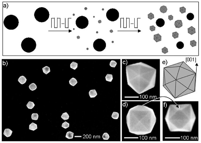
Nanocrystals are fundamental to modern science and technology. Mastery over the shape of a nanocrystal enables control of its properties and enhancement of its usefulness for a given application. Our aim is to present a comprehensive review of current research activities that center on the shape-controlled synthesis of metal nanocrystals. We begin with a brief introduction to nucleation and growth within the context of metal nanocrystal synthesis, followed by a discussion of the possible shapes that a metal nanocrystal might take under different conditions. We then focus on a variety of experimental parameters that have been explored to manipulate the nucleation and growth of metal nanocrystals in solution-phase syntheses in an effort to generate specific shapes. We then elaborate on these approaches by selecting examples in which there is already reasonable understanding for the observed shape control or at least the protocols have proven to be reproducible and controllable. Finally, we highlight a number of applications that have been enabled and/or enhanced by the shape-controlled synthesis of metal nanocrystals. We conclude this article with personal perspectives on the directions toward which future research in this field might take.


Figures

Figure 1
Figure 1
Plot of atomic concentration against time, illustrating the generation of atoms, nucleation, and subsequent growth (modified with permission from [33], copyright 1950 American Chemical Society).
Figure 2
Figure 2
Snapshots from a first-principles molecular dynamics simulation showing the reaction of a PtCl2(H2O)2 complex with a Pt12Cl4 cluster. Pt: yellow. Cl: green. O: red. H: white. Simulation time: a) 0.0 ps, b) 0.6 ps, c) 1.3 ps, d) 2.0 ps, e) 3.2 ps, and f) 5.0 ps (modified with permission from [35d], copyright 2003 American Chemical Society).
Figure 3
Figure 3
a) Positive-mode mass spectrum of a freshly prepared 1 mM aqueous AgNO3 solution. Note that Cs+ was added in the form of CsNO3 as a reference for concentration calibration. b) Plots of the concentrations of different silver species versus time, when a 1 mM aqueous AgNO3 solution was aged in air (modified with permission from [37], copyright 2007 Wiley-VCH).
Figure 4
Figure 4
Idealized representation of full-shell metal clusters with “magic numbers” of atoms, which are built upon the densest sphere packing (modified with permission from [49c], copyright 1999 Elsevier).
Figure 5
Figure 5
A schematic illustration of the reaction pathways that lead to fcc metal nanocrystals having different shapes. First, a precursor is reduced or decomposed to form the nuclei (small clusters). Once the nuclei have grown past a certain size, they become seeds with a single-crystal, singly twinned, or multiply twinned structure. If stacking faults are introduced, the plate-like seeds will be formed. The green, orange, and purple colors represent the {100}, {111}, and {110} facets, respectively. Twin planes are delineated in the drawing with red lines. The parameter R is defined as the ratio between the growth rates along the 〈100〉 and 〈111〉 directions (modified with permission from [26], copyright 2007 Wiley-VCH).
Figure 6
Figure 6
a) Schematic of a decahedron, which can be considered as the assembly of five single-crystal, tetrahedral units sharing a common edge. Since the theoretical angle between two {111} planes of a tetrahedron is 70.53°, five tetrahedrons joined with {111} twin planes will leave a gap of 7.35°. b, c) High-resolution TEM images of a decahedral Ag nanocrystal (modified with permission from [58b], copyright 2007 Royal Society of Chemistry). d) Schematic of a plate-like seed with a random hexagonal close-packed (rhcp) structure. Note that stacking faults and/or lamellar twins are introduced into the crystal lattice. e, f) High-resolution TEM images taken from the side face of a Ag nanoplate (modified with permission from [58b], copyright 2007 Royal Society of Chemistry).
Figure 7
Figure 7
Details of a polyol synthesis of Ag nanocrystals in which AgNO3 and PVP serve as the Ag precursor and capping agent, respectively. The reaction was performed in air and 0.06 mM NaCl was added. Reaction times: a, b) 10 min, c, d) 2 h, and e, f) 44 h. a, c, e) Photographs of the reaction solution, in which the yellow color indicates the presence of Ag nanocrystals. b, d, f) TEM images of the Ag nanocrystals produced at each time. Single-crystal and twinned nanocrystals are labeled as sc and tw, respectively. As the reaction proceeded, the twinned nanocrystals were removed due to oxidative etching, but the single-crystal species remained and accumulated in the solution because of their higher resistance to oxidative etching (modified with permission from [62], copyright 2004 American Chemical Society).
Figure 8
Figure 8
a) A schematic model of a hexagonal plate with a single twin plane, which contains concave-type (A) and convex-type (B) faces. The concave-type surface serves as the primary site for atomic addition, facilitating the transformation of hexagonal plates into triangular plates. b) Schematic models of a decahedron seed and a five-fold twinned rod. The five variants in the decahedron are separated by {111}-type twin planes. In the five-fold twinned rod, the side faces are {100} and the end faces are {111} faces. The green and orange colors represent the {100} and {111} facets, respectively. Twin planes are delineated in the figure with red lines (modified with permission from [52b], copyright 2005 Wiley-VCH).
Figure 9
Figure 9
Shape evolution during the successive stages of growth for an imaginary 2-D crystal. a) Rapid addition to the y-edges (relative to the x-edges) results in the elongation of the x-edges and the eventual disappearance of the y-edges, and b) vice versa. The length of arrow is directly proportional to the growth rate. When the crystal is enclosed by a single set of planes, the shape will become stable over growth time unless the surface is modified again due to etching, Ostwald ripening, and/or capping.
Figure 10
Figure 10
A schematic illustration of the overgrowth process of Ag nanocrystals, in which Ag atoms are continuously deposited onto the {100} facets of a Ag nanocube to eventually result in an octahedron enclosed by {111} facets (modified with permission from [67a], copyright 2006 Wiley-VCH).
Figure 11
Figure 11
Electron microscopy characterization of the binary Pt/Pd core-shell nanocrystals obtained through the heteroepitaxial deposition of Pd on cubic Pt seeds: a, b) cube; c, d) cuboctahedron; and e, f) octahedron (modified with permission from [94a], copyright 2007 Nature Publishing Group).
Figure 12
Figure 12
Electron microscopy characterization of the binary Pd/Pt core-shell nanoplates obtained through the heteroepitaxial growth of Pt shells on Pd nanoplate seeds. a-d) STEM images and the corresponding cross-sectional compositional line profiles of Pd/Pt core-shell nanoplates for a, b) hexagonal and c, d) triangular plates. e, f) HRTEM images taken from the side faces of the Pd/Pt core-shell nanoplates, which show the continuous lattice fringes from the Pd core (lattice spacing: 2.22 Å) to the Pt shell (lattice spacing: 2.29 Å) (modified with permission from [94c], copyright 2008 American Chemical Society).
Figure 13
Figure 13
Electron microscopy images of single-crystal Pd nanocrystals: a) Wulff polyhedrons prepared in ethylene glycol with PVP as a capping agent (modified with permission from [64a], copyright 2005 American Chemical Society); b) slightly truncated nanocubes prepared in ethylene glycol with PVP as a capping agent and FeIII species as an etchant (modified with permission from [78b], copyright 2005 American Chemical Society); c) nanocubes prepared with PVP as a reductant in water and in the presence of KBr; d) nanobars prepared in a mixture of 90.9% water and 9.1% ethylene glycol, in the presence of KBr; e) nanorods prepared in a mixture of 72.7% ethylene glycol and 27.3% water, in the presence of KBr (modified with permission from [64d], copyright 2007 American Chemical Society); and f) octahedrons prepared with citric acid as a reducing agent and capping agent at a high concentration of Pd precursor (modified with permission from [80], copyright 2007 Wiley-VCH).
Figure 14
Figure 14
Electron microscopy images of Pd nanocrystals with twin defects: a) decahedrons prepared with citric acid as a reducing agent and capping agent at high concentrations for both Pd precursor and citric acid (modified with permission from [80], copyright 2007 Wiley-VCH); b) icosahedrons prepared with citric acid as a reducing agent and capping agent at a low concentration of Pd precursor (modified with permission from [80], copyright 2007 Wiley-VCH); c) five-fold twinned nanorods (as a mixture with cubebs) prepared with ascorbic acid as a reductant in water and in the presence of bromide; d) single-twinned right bipyramids (as a mixture with cubes) prepared with ascorbic acid as a reductant in water and in the presence of bromide (modified with permission from [68], copyright 2007 Elsevier); e) triangular nanoplates prepared in ethylene glycol and in the presence of FeCl3 and HCl (modified with permission from [60], copyright 2005 American Chemical Society); and f) hexagonal nanoplates prepared with PVP as a reductant in water (modified with permission from [59b], copyright 2006 American Chemical Society).
Figure 15
Figure 15
Electron microscopy images of single-crystal Ag nanocrystals: a) cuboctahedrons prepared in ethylene glycol with PVP as a capping agent (modified with permission from [62], copyright 2004 American Chemical Society); b) nanocubes prepared in ethylene glycol with PVP as a capping agent (modified with permission from [78a], copyright 2002 American Association for the Advancement of Science); c) truncated octahedrons prepared in 1,5-pentanediol in the presence of PVP and Cu2+ ions; d) octahedrons prepared in 1,5-pentanediol in the presence of PVP and Cu2+ ions (modified with permission from [67a], copyright 2006 Wiley-VCH); e) nanocubes prepared by a modified silver mirror reaction in the presence of Br- with glucose as a reducing agent (modified with permission from [110b], copyright 2005 American Chemical Society); and f) nanobars prepared in ethylene glycol in the presence of PVP and Br- (modified with permission from [63b], copyright 2007 American Chemical Society).
Figure 16
Figure 16
Electron microscopy images of Ag nanocrystals with twin defects: a) singly twinned right bipyramids prepared in ethylene glycol in the presence of PVP and Br- (modified with permission from [63a], copyright 2006 American Chemical Society); b) singly twinned nanobeams prepared in ethylene glycol in the presence of PVP and Br- (modified with permission from [69], copyright 2006 American Chemical Society); c) five-fold twinned nanorods prepare in ethylene glycol with PVP as a capping agent (modified with permission from [65], copyright 2005 American Chemical Society); d) nanoplates prepared via the light-induced conversion of Ag nanospheres (modified with permission from [115a], copyright 2003 Nature Publishing Group); e) nanoplates prepared in water with PVP as a reductant (modified with permission from [59a], copyright 2006 Wiley-VCH); and f) nanobelts formed by refluxing an aqueous dispersion of Ag colloids (modified with permission from [61b], copyright 2003 American Chemical Society).
Figure 17
Figure 17
Electron microscopy images of Au nanocrystals: a) octahedrons prepared with PVP as a capping agent in polyethylene glycol 600 (PEG 600) (modified with permission from [120b], copyright 2007 Wiley-VCH); b) truncated tetrahedrons prepared with PVP as a capping agent in tetraethylene glycol (modified with permission from [120e], copyright 2008 American Chemical Society); c) icosahedrons prepared in ethylene glycol with a low concentration of Au precursor (modified with permission from [91], copyright 2004 Wiley-VCH); d) decahedrons prepared in diethylene glycol with a high concentration of PVP (modified with permission from [120e], copyright 2008 American Chemical Society); e) truncated nanocubes prepared in 1,5-pentanediol in the presence of Ag+ ions with PVP as a capping agent; and f) nanocubes prepared in 1,5-pentanediol in the presence of Ag+ ions with PVP as a capping agent (modified with permission from [121b], copyright 2006 American Chemical Society).
Figure 18
Figure 18
Electron microscopy images of anisotropic Au nanocrystals: a) hexagonal nanoplates prepared with ortho-phenylenediamine as a reductant (modified with permission from [122b], copyright 2004 Wiley-VCH); b) triangular nanoplates prepared with Sargassum sp. as a reductant (modified with permission from [122i], copyright 2005 American Chemical Society); c) nanobelts prepared with ascorbic acid as a reducing agent and SDSn and CTAB as surfactants (modified with permission from [126], copyright 2008 American Chemical Society); and d) five-fold twinned nanorods prepared via a seeding process with CTAB as a capping agent (modified with permission from [79a], copyright 2001 American Chemical Society).
Figure 19
Figure 19
Electron microscopy images of various Au nanocrystals prepared via a seeding process: a) nanorods prepared in the presence of Au seeds and Ag+ ions and with CTAB as a capping agent (modified with permission from [128d], copyright 2005 American Chemical Society); b) multipods formed through an overgrowth mechanism (modified with permission from [131a], copyright 2004 American Chemical Society); c) dog bone-shaped nanocrystals formed through an overgrowth mechanism (modified with permission from [128d], copyright 2005 American Chemical Society); and d) dumbbell-shaped nanocrystals formed through an overgrowth mechanism (modified with permission from [131c], copyright 2005 Wiley-VCH).
Figure 20
Figure 20
a) A schematic illustrating the formation of a polymeric strand of oleylamine-AuCl complex; b) TEM image of ultrathin Au nanowires with an average diameter of 1.8 nm obtained by reducing the oleylamine-AuCl complex with 10-nm Ag nanoparticles in hexane; and c) HRTEM image showing <111> growth direction for most of the nanowires (modified with permission from [134a], copyright 2008 American Chemical Society).
Figure 21
Figure 21
Electron microscopy images of Pt nanocrystals with different shapes: a) nanocubes prepared in the presence of TTAB with a high concentration of NaBH4; b) cuboctahedrons prepared in the presence of TTAB with a low concentration of NaBH4 (modified with permission from [144a], copyright 2007 American Chemical Society); c) nanodendrites prepared with ascorbic acid as a reducing agent (modified with permission from [143b], copyright 2004 American Chemical Society); d) tetrapods prepared via a polyol process using FeIII species to reduce the number of seeds at the nucleation stage (modified with permission from [135b], copyright 2005 Wiley-VCH); e) nanowires prepared via a polyol process using FeIII species to slow down the reduction (modified with permission from [135a], copyright 2004 American Chemical Society); and f) nanowire network prepared in a two-phase water-chloroform system in the presence of CTAB with NaBH4 as a reducing agent (modified with permission from [144b], copyright 2007 American Chemical Society).
Figure 22
Figure 22
a) A schematic illustrating the electrochemical preparation of Pt tetrahexahedrons (THHs) from nanospheres: under the influence of the square-wave potential, Pt THHs can nucleate and grow at the expense of spherical particles; b) low-magnification SEM image of the Pt THHs; c, d) high-magnification SEM images of a Pt THH along different orientations, clearly showing the shape of the THH; e) geometrical model of an ideal THH; and f) high-magnification SEM image of a Pt THH, showing the imperfect vertices as a result of unequally sized neighboring facets (modified with permission from [145], copyright 2007 American Association for the Advancement of Science).
Figure 23
Figure 23
Schematic of Pt nanocrystals formed from seeds with different numbers of twin planes: a) zero, b) single, c) five, and d) multiple. Twin planes are delineated in the figure with red lines. Scale bars are 20 nm if not labeled in the images (modified with permission from [149], copyright 2007 American Chemical Society).
Figure 24
Figure 24
a) TEM image of five-fold twinned Cu nanorods prepared by reducing copper(II) bis(2-ethylhexyl)sulfosuccinate (Cu(AOT)2) with hydrazine in a mixture of isooctane and water (modified with permission from [152a], copyright 1997 American Chemical Society). b) SEM image of Cu nanowires with a circular cross-section prepared by reducing Cu(NO3)2 with hydrazine in the presence of sodium hydroxide and ethylenediamine [158].
Figure 25
Figure 25
TEM images of Rh nanocrystals with different shapes: a) nanocubes prepared via a polyol process at 190 °C (modified with permission from [164a], copyright 2005 American Chemical Society); b) multipods prepared via a polyol process at 140 °C (modified with permission from [64c], copyright 2006 Wiley-VCH); c) tetrahedrons prepared through the decomposition of Rh2(CO)4Cl2; and d) nanorods prepared through the decomposition of Rh(C5H8O2)3 (modified with permission from [164d], copyright 2007 Wiley-VCH).
Figure 26
Figure 26
Electron microscopy images of Fe, Co and Ni nanocrystals with different shapes: a) α-Fe nanoparticles prepared by thermal decomposition of Fe(CO)5 (modified with permission from [168f], copyright 2005 American Chemical Society); b) Fe nanocubes prepared by thermal decomposition of Fe[N(SiMe3)2]2 (modified with permission from [169a], copyright 2004 American Association for the Advancement of Science); c) ε-Co nanoparticles prepared by reducing CoCl2 with LiBEt3H (modified with permission from [171b], copyright 1999 American Institute of Physics); d) hcp-Co nanodisks prepared by rapid decomposition of Co2(CO)8 (modified with permission from [171h], copyright 2002 American Chemical Society); e) hcp-Co nanorods prepared by decomposition of [Co(η3-C8H13)(η4-C8H12)] (modified with permission from [173b], copyright 2003 Wiley-VCH); and f) fcc-Ni nanoplates prepared by decomposition of Ni(COD)2 in the presence of Fe(CO)5 (modified with permission from [176], copyright 2006 Institute of Physics Publishing).
Figure 27
Figure 27
Electron microscopy images of Bi nanocrystals with different shapes: a) nanospheres prepared using a microemulsion-based approach (modified with permission from [178a], copyright 2006 Wiley-VCH); b) nanocubes, c) triangular nanoplates, and d) nanobelts prepared via hydrothermal routes (modified with permission from [183e], copyright 2006 American Chemical Society).
Figure 28
Figure 28
a) SEM image of Pb nanowires prepared by a polyol process in the presence of PVP; the inset shows the cross-section of a broken nanowire; b) schematic illustrating the growth of a Pb nanowire with three components: root, stem, and tip. The Pb nanodroplets in the solution phase serve as the source of atoms for growth; c) TEM image of an individual Pb nanowire and the SAED patterns (insets) taken from its root and stem, respectively; and d) SEM image of Pb nanoplates prepared by increasing the concentration of PVP (modified with permission from [186b], copyright 2004 American Chemical Society).
Figure 29
Figure 29
a) Schematic illustrating the unit cell of chemically disordered fcc- and chemically ordered fct-FePt (modified with permission from [6b], copyright 2006 Wiley-VCH); b) TEM image of truncated fcc-FePt nanocubes prepared by simultaneous reduction of Pt(acac)2 and thermal decomposition of Fe(CO)5 (modified with permission from [97], copyright 2000 American Association for the Advancement of Science); and c) TEM image of Pt@Fe2O3 core-shell nanostructures prepare via a two-step process (modified with permission from [193f], copyright 2003 American Chemical Society).
Figure 30
Figure 30
Electron microscopy images of metal nanocrystals after aging: a) Ag nanocubes aged at 160 °C for 5 min in an ethylene glycol solution containing 1 mM HCl in the presence of 0.1 mM PVP (modified with permission from [197], copyright 2007 American Chemical Society); b) Ag nanoplates aged in water for 1 month at room temperature (modified with permission from [58b], copyright 2007 Royal Society of Chemistry); c) Pd nanocubes and d) Pd single-crystal nanorods aged in the reaction solution for 4 weeks at room temperature (modified with permission from [64d], copyright 2007 American Chemical Society). The insets in panel (a-d) show TEM images of the corresponding nanocrystals before the aging process, at the same magnification as in (a-d).
Figure 31
Figure 31
Calculated UV-visible extinction (black), absorption (red), and scattering (blue) spectra of Ag nanocrystals, illustrating the effect of shape on spectral characteristics: a) sphere, b) cube, c) tetrahedron, d) octahedron, e) triangular plate, and f) circular plate (modified with permission from [16e], copyright 2006 American Chemical Society).
Figure 32
Figure 32
a) SEM images of individual Ag nanobars and the corresponding normalized LSPR spectra. The longitudinal plasmon peak red-shifts with increasing aspect ratio for the nanobars. b) LSPR (scattering) spectra calculated using the DDA method for Ag nanobars 100, 150, and 200 nm in length, keeping width = 55 nm and height = 50 nm. c) SEM images of individual nanorice with the corresponding normalized LSPR spectra. d) Plot of longitudinal plasmon peak location versus aspect ratio. The peaks of both nanobars and nanorice red-shift with increasing length, but on average the peaks of nanobars are 80 nm red-shifted from nanorice (modified with permission from [63b], copyright 2007 American Chemical Society).
Figure 33
Figure 33
The normalized SERS spectra of 1,4-BDT adsorbed on a Ag nanocube with sharp corners (left panel, a-c) and a highly truncated Ag nanocube (right panel, d-f), at various angles relative to the polarization of the excitation laser. Each SEM image shows the nanocube used and the arrows indicate the polarization directions of incident laser. The scale bar applies to both images. The broad peak at 900-1000 cm-1 from the underlying silicon substrate was used as the reference for normalization (modified with permission from [207], copyright 2007 American Chemical Society).
Figure 34
Figure 34
Reflectance spectra obtained from crystalline lattices of a) Pb and b) Pb@SiO2 nanospheres with the incident light oriented perpendicular to their (111) planes. The insets in both figures show the corresponding top-view SEM images for the two crystalline lattices. Both scale bars in the insets are 200 nm (modified with permission from [178d], copyright 2006 Wiley-VCH).
Figure 35
Figure 35
SEM images of structures self-assembled from Ag nanocubes. The nanocubes were selectively functionalized with hydrophilic and hydrophobic thiolate SAMs and then allowed to assemble in water. The number of faces on each nanocube that were rendered hydrophobic is indicated in grey color in the bottom right corner of each panel, the remaining faces on the nanocube were hydrophilic. All nanocubes used in this study had a mean edge length of 97 ± 6 nm, as determined from 123 cubes (modified with permission from [226], copyright 2008 Wiley-VCH).

References

    1. Fahlman BD. Materials Chemistry. Vol. 1. Springer; Mount Pleasant, MI: 2007. pp. 282–283.
    1. See, for example, Halperin WP. Rev Mod Phys. 1986;58:533.Kreibig U, Vollmer M. Optical Properties of Metal Clusters. Springer; New York: 1995. http://esi-topics.com/nanocrystals/

    1. Reviews: Bawendi MG, Steigerwald ML, Brus LE. Annu Rev Phys Chem. 1990;41:477.Weller H. Adv Mater. 1993;5:88.Alivisatos AP. Science. 1996;271:933.Murray CB, Kagan CR, Bawendi MG. Annu Rev Mater Sci. 2000;30:545.

    1. See, for example, Likharev KK. IBM J Res Develop. 1988;32:144.Maheshwari V, Kane J, Saraf R. Adv Mater. 2008;20:284.

    1. See, for example, Mott NF. Rev Mod Phys. 1968;40:677.Markovich G, Collier CP, Henrichs SE, Remacle F, Levine RD, Heath JR. Acc Chem Res. 1999;32:415.

Publication types

LinkOut - more resources