Comparative Study
. 2008 Dec 16;47(50):13354-61.
doi: 10.1021/bi8017762.

Structure of a mutant form of proliferating cell nuclear antigen that blocks translesion DNA synthesis

Affiliations
Comparative Study

Structure of a mutant form of proliferating cell nuclear antigen that blocks translesion DNA synthesis

Bret D Freudenthal et al. Biochemistry. .

Abstract

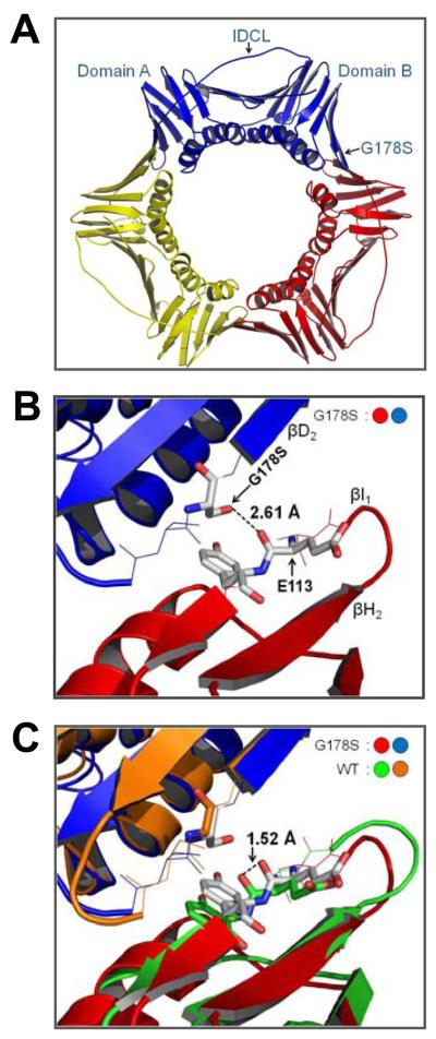
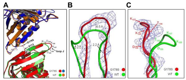
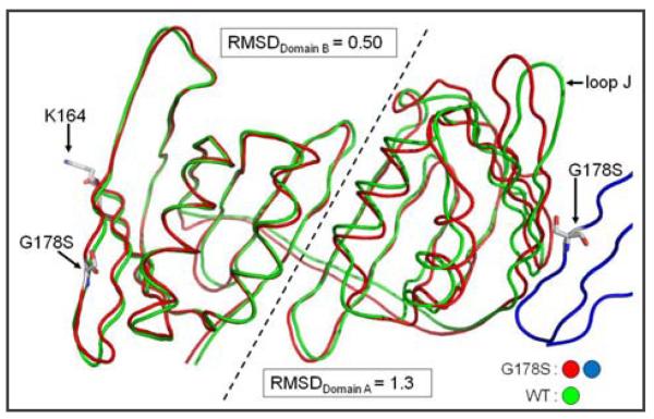
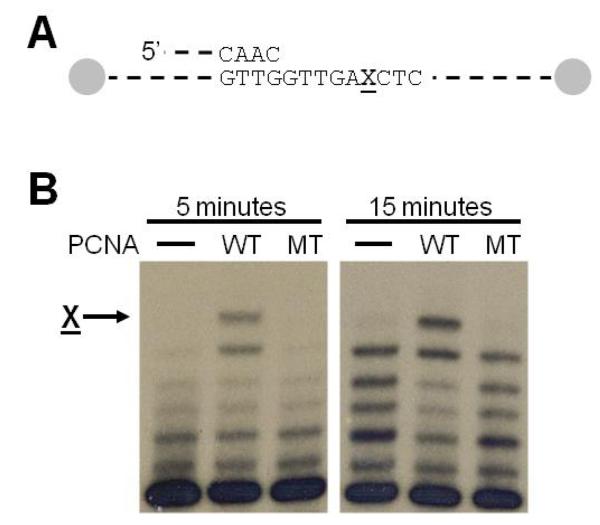
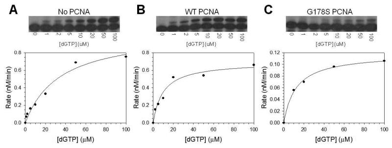
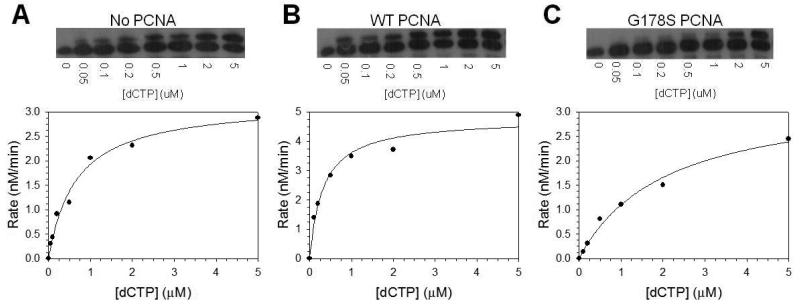
Proliferating cell nuclear antigen (PCNA) is a homotrimeric protein that functions as a sliding clamp during DNA replication. Several mutant forms of PCNA that block translesion DNA synthesis have been identified in genetic studies in yeast. One such mutant protein (encoded by the rev6-1 allele) is a glycine to serine substitution at residue 178, located at the subunit interface of PCNA. To improve our understanding of how this substitution interferes with translesion synthesis, we have determined the X-ray crystal structure of the PCNA G178S mutant protein. This substitution has little effect on the structure of the domain in which the substitution occurs. Instead, significant, local structural changes are observed in the adjacent subunit. The most notable difference between mutant and wild-type structures is in a single, extended loop (comprising amino acid residues 105-110), which we call loop J. In the mutant protein structure, loop J adopts a very different conformation in which the atoms of the protein backbone have moved by as much as 6.5 A from their positions in the wild-type structure. To improve our understanding of the functional consequences of this structural change, we have examined the ability of this mutant protein to stimulate nucleotide incorporation by DNA polymerase eta (pol eta). Steady state kinetic studies show that while wild-type PCNA stimulates incorporation by pol eta opposite an abasic site, the mutant PCNA protein actually inhibits incorporation opposite this DNA lesion. These results show that the position of loop J in PCNA plays an essential role in facilitating translesion synthesis.


Figures

Fig. 1
Fig. 1
Structure of the PCNA G178S Mutant Protein. (A) The trimeric form of the protein is shown with monomeric subunits in red, yellow, and blue. The inter-domain connecting loop (IDCL), domains A and B, and the G178S substitution are indicated on one of the subunits. (B) Close up, side view of the subunit interface with the Ser-178 substitution of the blue monomer and Tyr-114 and Glu-113 of the red monomer shown in stick format. The distance between the hydroxyl of Ser-178 and the backbone carbonyl of Glu-113 is indicated. (C) The superimposition of the structures of the G178S PCNA mutant protein and wild type PCNA (1PLQ). The distance between backbone carbonyl of Glu-113 in the wild type and mutant PCNA structures is indicated.
Fig. 2
Fig. 2
Conformation of loop J in the wild type and mutant structure. (A) Superimposition of the wild type and G178S PCNA mutant protein structures is shown with the Ser-178 substitution and Glu-113 represented in stick format and the hydrogen bond between them shown as black dots. The amino acid residues of loop J are indicated. (B) Close up view of loop J showing the electron density (level=2.0) for the G178S PCNA mutant protein and the backbone of the wild type and mutant proteins in ribbon representation. The distances between the wild type and mutant protein backbone are specified. (C) Side view of loop J with the position of the amino acid residues indicated.
Fig. 3
Fig. 3
Superimposition of the PCNA monomer backbone of wild type and mutant PCNA proteins. The monomeric subunit is lying on its side with the inter-domain connector loop in the back to allow the separate domains to be easily viewed. The adjacent mutant monomeric subunit is shown in blue with the G178S substitution indicated. The G178S substitution, the site of mono-ubiquitination (Lys-164), and loop J are indicated. Domains A and B of the monomeric subunit are separated by a dashed line and the RMSD values were independently determined for each domain.
Fig. 4
Fig. 4
Running start experiment with pol η on an abasic site. (A) Schematic diagram of the 31/68-mer substrate used in the running start assays with the ends of the template strand containing biotinstreptavidin blocks. The X indicates the location of the abasic site (B) Autoradiograph of the synthesis products after five or fifteen minutes following the addition of pol η. The arrow indicates incorporation opposite the abasic site. Lanes labeled WT contain the wild-type PCNA protein, and lanes labeled MT contain the G178S mutant PCNA protein.
Fig. 5
Fig. 5
Steady state kinetics of pol η on an abasic site. The rate of nucleotide incorporation was graphed as a function of dGTP concentration for (A) pol η alone, (B) pol η with wild type PCNA protein, (C) and pol η with the G178S PCNA mutant protein. Autoradiographs of the gels showing the incorporation of a single dGTP across from an abasic site at the indicated concentrations of nucleotide are shown above each graph. The solid lines represent the best fits of the data to the Michaelis–Menten equation, and the Vmax and Km steady state parameters are given in Table 2.
Fig. 6
Fig. 6
Steady state kinetics of pol η on non-damaged DNA. The rate of nucleotide incorporation was graphed as a function of dCTP concentration for (A) pol η alone, (B) pol η with wild type PCNA protein, (C) and pol η with the G178S PCNA mutant protein. Autoradiographs of the gel showing the single incorporation of dCTP at the indicated concentrations of nucleotide are shown above each graph. The solid lines represent the best fits of the data to the Michaelis–Menten equation, and the Vmax and Km steady-state parameters are given in Table 2.

References

    1. Goodman MF. Error-prone repair DNA polymerases in prokaryotes and eukaryotes. Annu. Rev. Biochem. 2002;71:17–50. - PubMed
    1. Prakash S, Prakash L. Translesion DNA synthesis in eukaryotes: a one- or two-polymerase affair. Genes Dev. 2002;16:1872–1883. - PubMed
    1. Prakash S, Johnson RE, Prakash L. Eukaryotic translesion synthesis DNA polymerases: specificity of structure and function. Annu. Rev. Biochem. 2005;74:317–353. - PubMed
    1. Johnson RE, Prakash S, Prakash L. Efficient bypass of a thymine-thymine dimer by yeast DNA polymerase, Pol eta. Science. 1999;283:1001–1004. - PubMed
    1. Washington MT, Johnson RE, Prakash S, Prakash L. Accuracy of thymine-thymine dimer bypass by Saccharomyces cerevisiae DNA polymerase eta. Proc. Natl. Acad. Sci U.S.A. 2000;97:3094–3099. - PMC - PubMed

Publication types

MeSH terms

Substances

LinkOut - more resources