. 2008 Dec 23;47(51):13554-63.
doi: 10.1021/bi801610c.

Structural studies of human histone deacetylase 8 and its site-specific variants complexed with substrate and inhibitors

Affiliations

Structural studies of human histone deacetylase 8 and its site-specific variants complexed with substrate and inhibitors

Daniel P Dowling et al. Biochemistry. .

Abstract

Metal-dependent histone deacetylases (HDACs) require Zn(2+) or Fe(2+) to regulate the acetylation of lysine residues in histones and other proteins in eukaryotic cells. Isozyme HDAC8 is perhaps the archetypical member of the class I HDAC family and serves as a paradigm for studying structure-function relationships. Here, we report the structures of HDAC8 complexes with trichostatin A and 3-(1-methyl-4-phenylacetyl-1H-2-pyrrolyl)-N-hydroxy-2-propenamide (APHA) in a new crystal form. The structure of the APHA complex reveals that the hydroxamate CO group accepts a hydrogen bond from Y306 but does not coordinate to Zn(2+) with favorable geometry, perhaps due to the constraints of its extended pi system. Additionally, since APHA binds to only two of the three protein molecules in the asymmetric unit of this complex, the structure of the third monomer represents the first structure of HDAC8 in the unliganded state. Comparison of unliganded and liganded structures illustrates ligand-induced conformational changes in the L2 loop that likely accompany substrate binding and catalysis. Furthermore, these structures, along with those of the D101N, D101E, D101A, and D101L variants, support the proposal that D101 is critical for the function of the L2 loop. However, amino acid substitutions for D101 can also trigger conformational changes of Y111 and W141 that perturb the substrate binding site. Finally, the structure of H143A HDAC8 complexed with an intact acetylated tetrapeptide substrate molecule confirms the importance of D101 for substrate binding and reveals how Y306 and the active site zinc ion together bind and activate the scissile amide linkage of acetyllysine.


Figures

Figure 1
Figure 1
Proposed mechanism of histone deacetylase 8 (HDAC8) (–, –25).
Figure 2
Figure 2
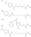
Molecular structures of trichostatin A (TSA), suberoylanilide hydroxamic acid (SAHA), 3-(1-methyl-4-phenylacetyl-1H-2-pyrrolyl)-N-hydroxy-2-propenamide (APHA), and 4-(dimethylamino)-N-[7-(hydroxyamino)-7-oxoheptyl]-benzamide (M344).
Figure 3
Figure 3
Packing of HDAC8 molecules in the new monoclinic crystal form. Monomers A, B, and C appear as pink, green, and dark blue, respectively. The unit cell is depicted in pink (a = 87.90 Å, b = 90.71 Å, c = 92.14 Å, β= 94.6°).
Figure 4
Figure 4
Simulated-annealing omit map of APHA (magenta, contoured at 3.5σ) and Zn2+ (cyan, contoured at 11σ) in monomer B of the HDAC8-APHA complex. α-Helices B4 and C (which contains R167) of monomer C are shown in light blue. The side chain of D101 in the L2 loop of monomer B accepts hydrogen bonds from the pyrrole C=O group of APHA and R167 in helix C of monomer C. Metal coordination and hydrogen bond interactions are shown as solid black and dotted red lines, respectively.
Figure 5
Figure 5
Superposition of unliganded monomer C (light blue) and APHA-complexed monomer B (light green) of HDAC8 (Zn+2 is grey). Inhibitor binding results in the ordering of the L2 loop, which appears to be mediated by an enzyme-inhibitor hydrogen bond with D101 (red dotted line, also illustrated in Figure 4). A second zinc binding site is observed in the ordered L2 loop of monomer A only. Zn2+B (grey sphere) interacts with H78 (2.2 Å), D87 (2.6 Å), H90 (2.0 Å), and D92 (2.6 Å).
Figure 6
Figure 6
Binding of the inhibitor M344 to D101E, D101A, D101N, and D101L HDAC8 variants. The two monomers of the crystallographic dimer are light green and light blue. Simulated annealing omit maps of inhibitor molecules and residue 101 are superimposed (contoured at 3σ, except for D101A, which is contoured at 2.5σ). Two conformations of the bound inhibitor are observed in the active sites of D101E, D101A and D101N HDACs (the “yellow” conformation is similar to that observed for binding to the wild-type enzyme). D101L HDAC8 crystallizes in a different space group so the orientation of monomers is different from that observed in the D101E, D101A, and D101N variants. Electron density in the active site of D101L HDAC8 is interpreted as a mixture of 50% bound inhibitor and 50% two zinc-bound solvent molecules that essentially coincide with the positions of the zinc-bound oxygen atoms of the inhibitor (inhibitors bound to each monomer are show in yellow and green, respectively). Zinc-bound solvent molecules in D101L HDAC8 are show in cyan and contoured at 6σ. Zinc and potassium ions are grey and purple, respectively. Zinc coordination interactions are indicated by solid black lines and hydrogen bonds are indicated by dotted red lines.
Figure 7
Figure 7
Square pyramidal Zn2+ coordination polyhedron in the unliganded form of D101L HDAC8: D178, D267, and two solvent molecules occupy equatorial positions, and H180 occupies the apical position (atoms are color coded as follows: carbon = light green, nitrogen = blue, oxygen = red, Zn2+ = grey sphere, H2O = red spheres). Metal coordination and hydrogen bond interactions are shown in solid black and dotted red lines, respectively. Superimposed are the coordinates of the Y306F HDAC8-substrate complex (PDB accession code 2V5W) (25), with atoms color coded similarly except that carbon = light blue, Zn2+ = cyan sphere, and H2O = pink sphere. Note the correspondence of nonprotein coordination sites on zinc.
Figure 8
Figure 8
Comparison of the wild-type HDAC8-M344 complex (PDB accession code 1T67, light blue protein, purple residues and inhibitor) and the D101L HDAC8-M344 complex (light green protein, green residues and inhibitor) illustrates the “in” and “out” conformations of the aromatic side chains of W141 and Y111. Note that when W141 adopts the “out” conformation, a disordered glycerol molecule (Gol) binds in the resulting void; when Y111 adopts the “out” conformation, the side chain of L31 moves to fill the resulting void.
Figure 9
Figure 9
(a) Simulated annealing omit map (contoured at 2σ) showing substrate binding to H143A HDAC8. Metal coordination and hydrogen bond interactions are shown as solid black and dotted red lines, respectively. (b) Superposition of the H143A HDAC8-substrate complex (tan) and the previously reported (25) Y306F HDAC8-substrate complex (light blue). The zinc ion and water molecules observed in the H143A HDAC8-substrate complex appear as grey and red spheres, respectively. The zinc ion and zinc-bound water molecule observed in the Y306F HDAC8-substrate complex appear as cyan and pink spheres, respectively. Metal coordination and hydrogen bond interactions are shown as solid black and dotted red lines for the H143A HDAC8-substrate complex only.

References

    1. Gilbert N, Ramsahoye B. The relationship between chromatin structure and transcriptional activity in mammalian genomes. Brief Funct Genomic Proteomic. 2005;4:129–142. - PubMed
    1. Holbert MA, Marmorstein R. Structure and activity of enzymes that remove histone modifications. Curr Opin Struct Biol. 2005;15:673–680. - PubMed
    1. Vaquero A, Loyola A, Reinberg D. The constantly changing face of chromatin. Sci Aging Knowledge Environ 2003. 2003:RE4. - PubMed
    1. Cosgrove MS, Wolberger C. How does the histone code work? Biochem Cell Biol. 2005;83:468–476. - PubMed
    1. Eberharter A, Becker PB. Histone acetylation: a switch between repressive and permissive chromatin. Second in review series on chromatin dynamics. EMBO Rep. 2002;3:224–229. - PMC - PubMed

Publication types