Review
. 2009 Jan;33(1):191-205.
doi: 10.1111/j.1574-6976.2008.00149.x. Epub 2008 Nov 27.

Global phenotypic characterization of bacteria

Affiliations
Review

Global phenotypic characterization of bacteria

Barry R Bochner. FEMS Microbiol Rev. 2009 Jan.

Abstract

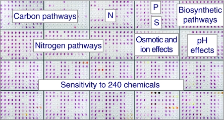
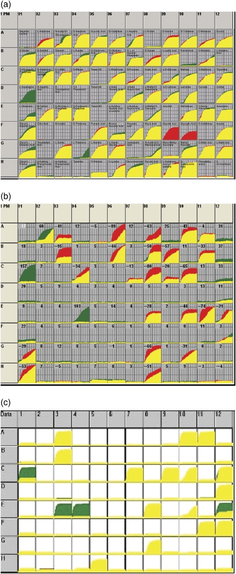
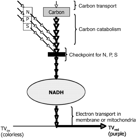
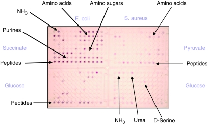
The measure of the quality of a systems biology model is how well it can reproduce and predict the behaviors of a biological system such as a microbial cell. In recent years, these models have been built up in layers, and each layer has been growing in sophistication and accuracy in parallel with a global data set to challenge and validate the models in predicting the content or activities of genes (genomics), proteins (proteomics), metabolites (metabolomics), and ultimately cell phenotypes (phenomics). This review focuses on the latter, the phenotypes of microbial cells. The development of Phenotype MicroArrays, which attempt to give a global view of cellular phenotypes, is described. In addition to their use in fleshing out and validating systems biology models, there are many other uses of this global phenotyping technology in basic and applied microbiology research, which are also described.


Figures

Fig. 1
Fig. 1
The 1920 phenotypic assays in the PM set for bacteria. PMs are sets of phenotypic assays performed in 96-well microplates. The microplate wells contain chemicals dried on the bottom to create unique culture conditions after rehydration. Assays are initiated by inoculating all wells with cell suspensions. After incubation, some of the wells turn various shades of purple due to reduction of a tetrazolium dye as the cells respire. The variable level of purple color indicates that the cells are metabolically active and respiring in some wells but not others. Other colors such as orange are the colors of other chemicals in the wells. Microplates in the PM set are organized into functional groups as labeled in the Figure. Assays of C, N, P, and S metabolism provide information about which metabolic pathways are present and active in the cells. Assays of ion, pH, and chemical sensitivities provide information on stress and repair pathways that are present and active in cells.
Fig. 3
Fig. 3
Using PM technology to detect changes in C metabolism. (a) An example of the compared C-source metabolic activities of two bacterial strains. Assays were performed in Biolog PM1 MicroPlates that contain a negative control well (well A1) and 95 different potential C-sources. Kinetic data were collected using the Biolog omnilog instrument and software. The curves show the time course (horizontal axis) of the amount of purple color formed from tetrazolium dye reduction (vertical axis) in each of the 96 wells. Data from one strain are shown in red, the other strain in green, and yellow is the overlapping of the two kinetic curves. C-sources more rapidly metabolized by the first strain are shown in red (F9, glycolic acid; F10, glyoxylic acid), and by the second strain are shown in green (D1, l-asparagine; G4, l-threonine), and metabolized equally are shown in yellow. (b) An example of the compared C-source metabolic activities of a strain of Yersinia pseudotuberculosis strain 15 478 when tested at two temperatures. The strain was assayed for C-metabolism using Biolog PM1 MicroPlates. For most C-sources, metabolism was more rapid at the warmer temperature. However, compared with the warmer temperature of 33°C (shown in red) the strain showed increased metabolism of three C-sources at 26°C (shown in green: well A2, l-arabinose; C1, d-glucose-6-PO4; E4, d-fructose-6-PO4). (c) An example of the compared C-source metabolic activities of an isogenic pair of Listeria monocytogenes strains (P14 vs. P14 prfA*). The strains were assayed for their C- metabolism using Biolog PM1 MicroPlates. Compared with its wild-type parental strain (shown in red), the hyperpathogenic prfA* strain (shown in green) exhibited increased metabolism of hexose phosphates as C-sources (well C1, d-glucose-6-PO4; E3, d-glucose-1-PO4; E4, d-fructose-6-PO4).
Fig. 2
Fig. 2
The coordinated linkage of metabolic pathways. Schematic diagram of major metabolic pathways in bacteria and how their activities are converted to a colorimetric readout. A C-source that can be transported into a cell and metabolized to produce NADH will engender a redox potential and flow of electrons to reduce a tetrazolium dye (Bochner & Savageau, 1977) such as tetrazolium violet (TV), thereby producing purple color. The more rapid this metabolic flow, the more quickly purple color is formed. However many cells exhibit a phenomenon of checkpoint control, where the catabolism of the C-source is restricted if the cell does not also have sufficient levels of N, P, and S. This enables assays where one can also measure these N, P, and S catabolic pathways. The more active they are, the more rapid the catabolism of the C-source and the more quickly purple color is formed.
Fig. 4
Fig. 4
Regulation of N metabolism by C metabolism is different in Escherichia coli vs. Staphylococcus aureus. Escherichia coli and S. aureus change their N-source metabolism in very different ways in response to the C-source they have available. Assays were performed in Biolog PM3 MicroPlates, which contain a negative control well (well A1) and 95 different potential N-sources. Cells are inoculated in a suspending medium with different C-sources, such as glucose, pyruvate, and succinate, as indicated in the figure. In E. coli, glucose represses the activity of many N-catabolic pathways and the set of active pathways is a subset of the pathways active on succinate. By contrast, in S. aureus, glucose represses the activity of many N-catabolic pathways but it also activates an entirely different set of active pathways compared with pyruvate.

References

    1. Baba T, Huan HC, Datsenko K, Wanner BL, Mori H. The applications of systematic in-frame, single-gene knockout mutant collection of Escherichia coli K-12. Method Mol Biol. 2008;416:183–194. - PubMed
    1. Bailey AM, Paulsen IT, Piddock LJ. RamA confers multidrug resistance in Salmonella enterica via increased expression of acrB, which is inhibited by chlorpromazine. Antimicrob Agents Ch. 2008;52:3604–3611. - PMC - PubMed
    1. Bibb MJ. Regulation of secondary metabolism in streptomycetes. Curr Opin Microbiol. 2005;8:208–215. - PubMed
    1. Biswas S, Biswas I. Role of HtrA in surface protein expression and biofilm formation by Streptococcus mutants. Infect Immun. 2005;73:6923–6934. - PMC - PubMed
    1. Bochner B. Methods for detection, identification and speciation of Listerias. US Patent 5,134,063.

Publication types