. 2009 Feb;10(2):201-17.
doi: 10.1111/j.1600-0854.2008.00856.x. Epub 2008 Nov 18.

STAM adaptor proteins interact with COPII complexes and function in ER-to-Golgi trafficking

Affiliations

STAM adaptor proteins interact with COPII complexes and function in ER-to-Golgi trafficking

Neggy Rismanchi et al. Traffic. 2009 Feb.

Abstract

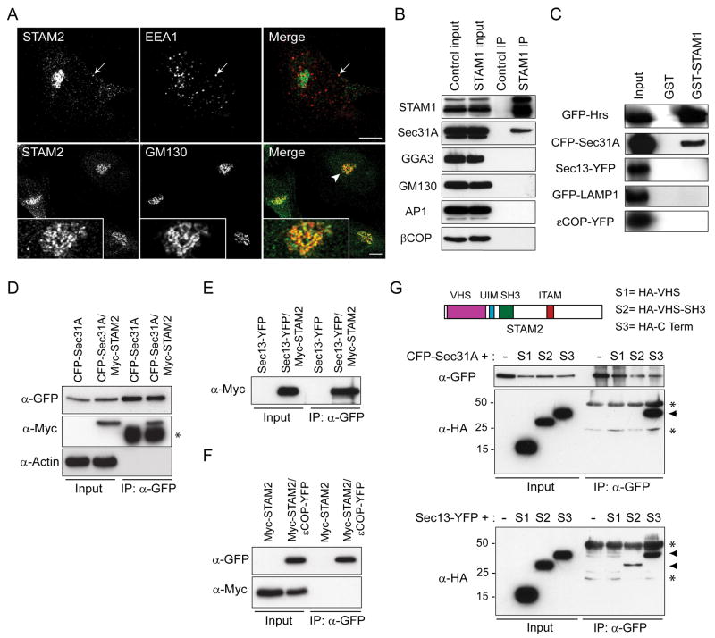
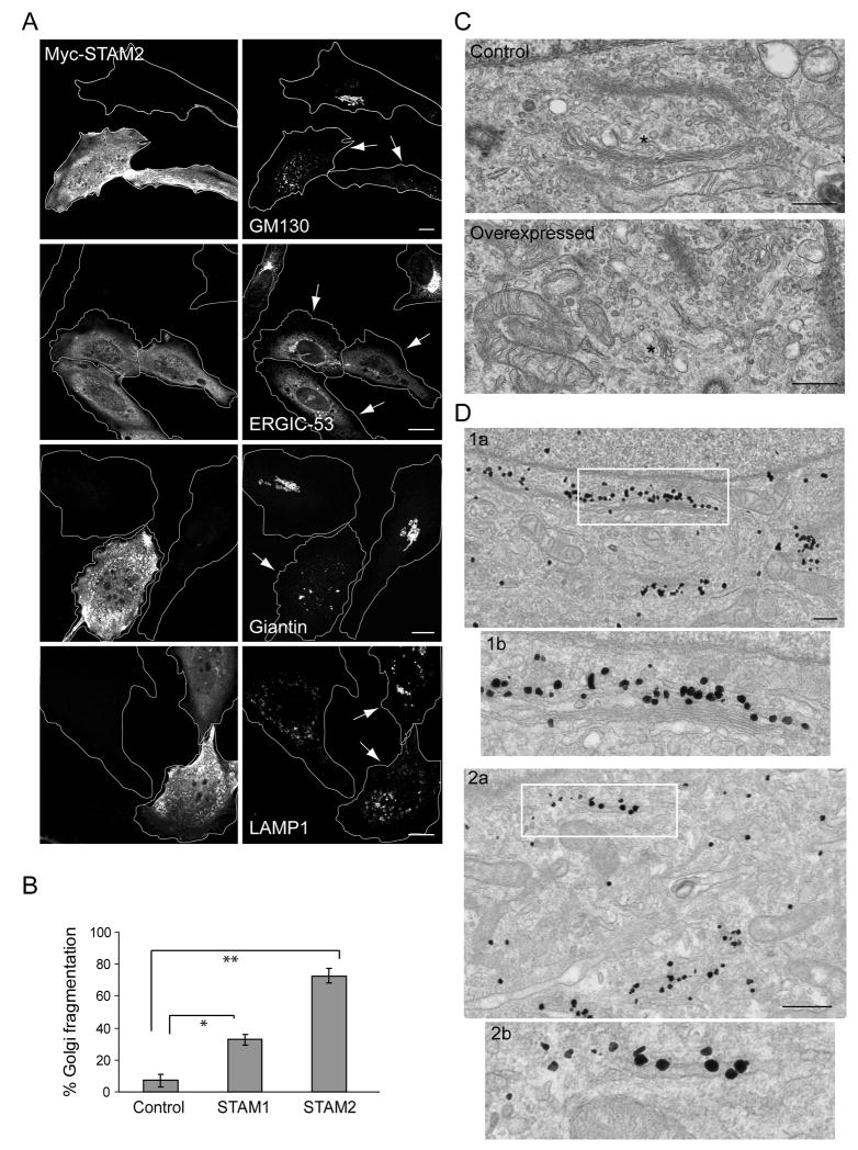
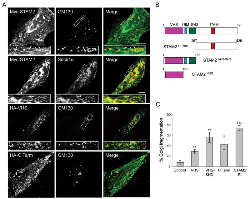
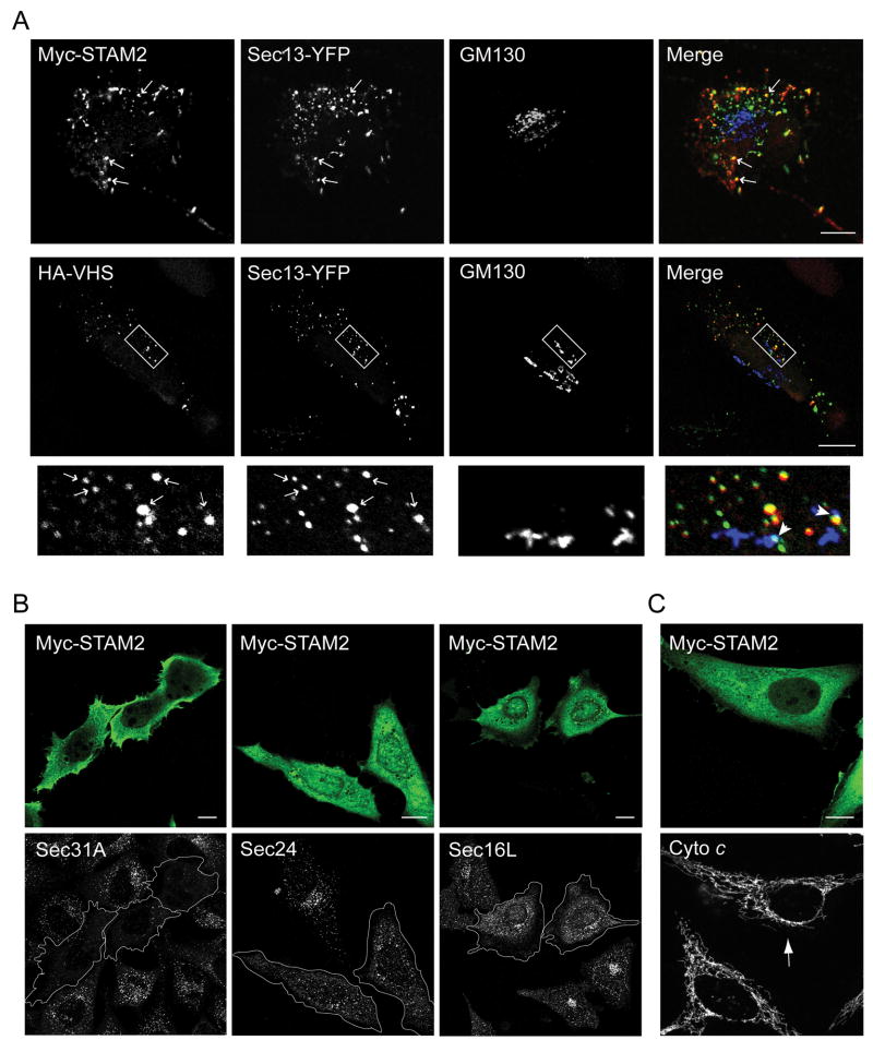
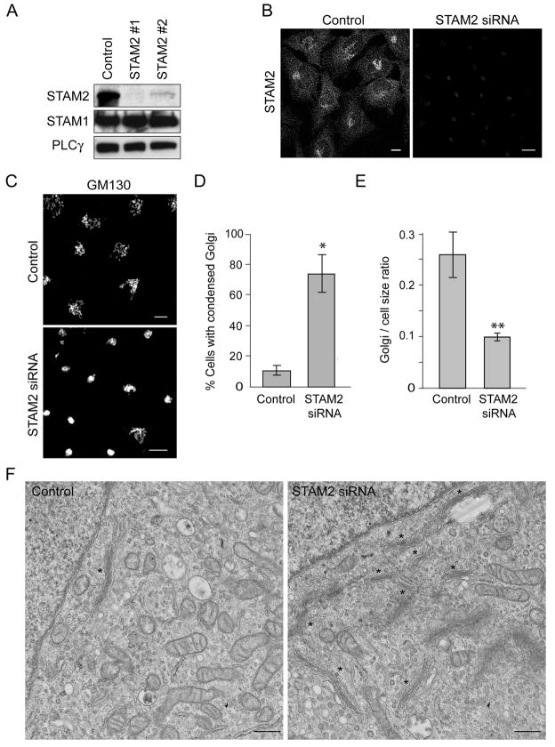
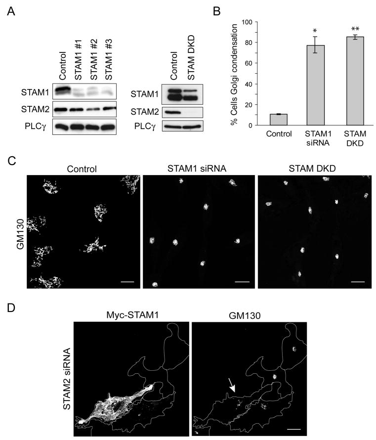
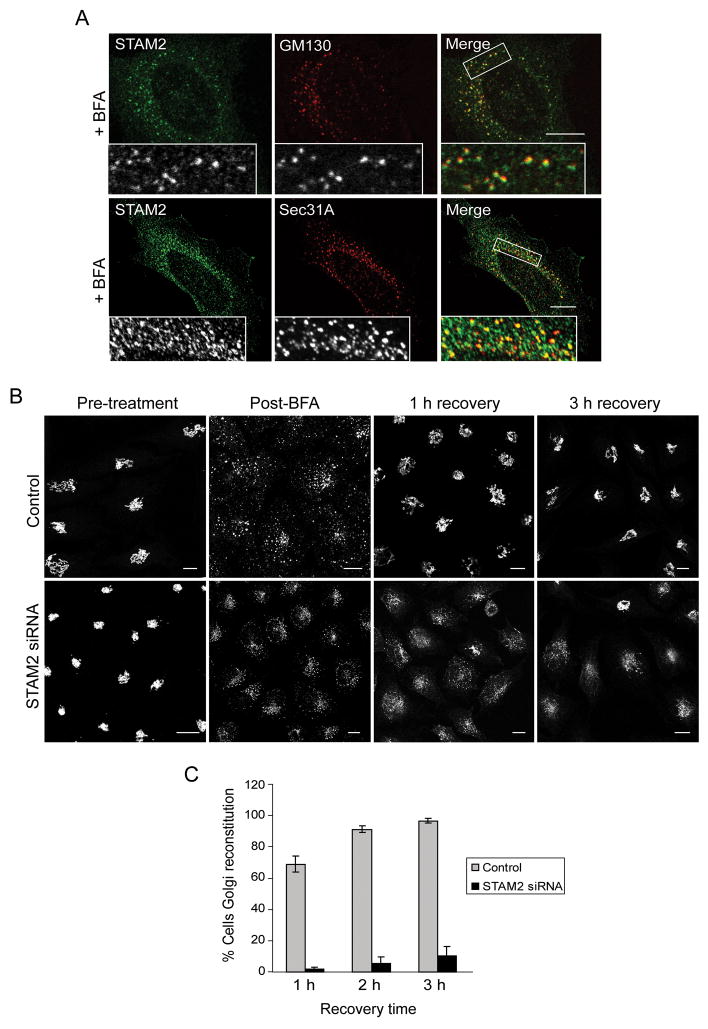
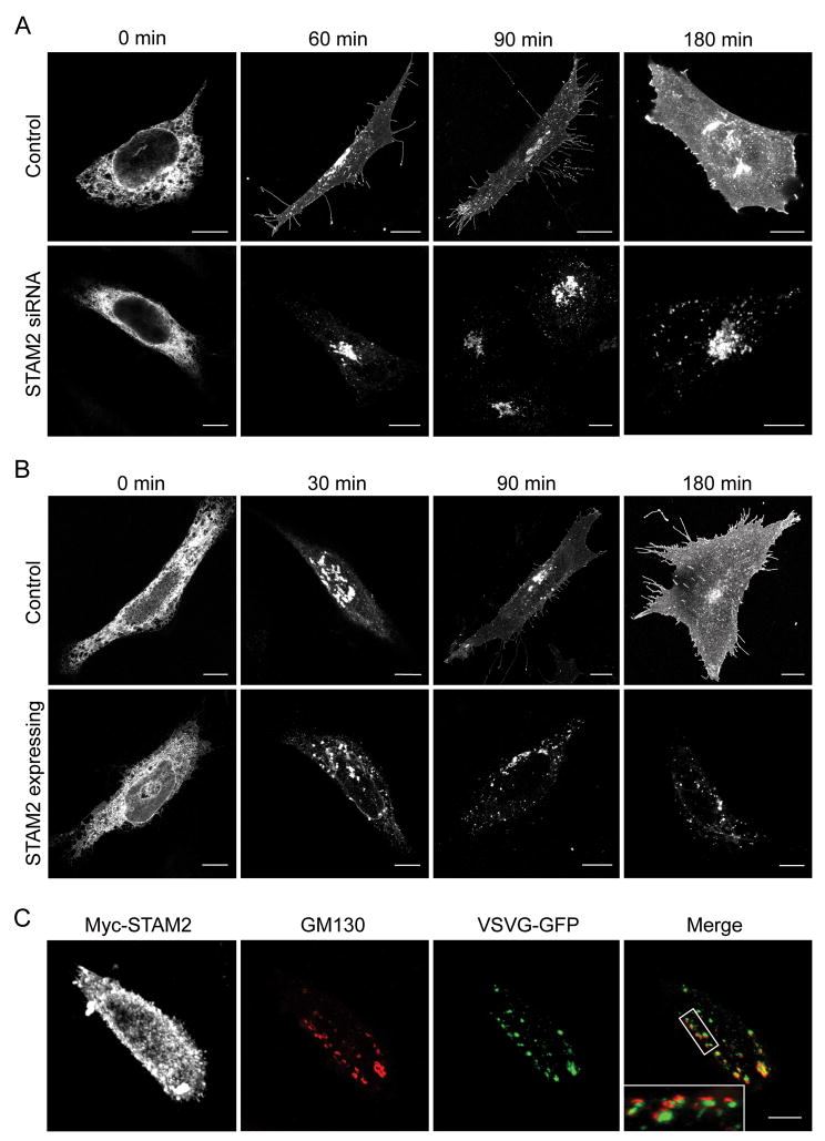
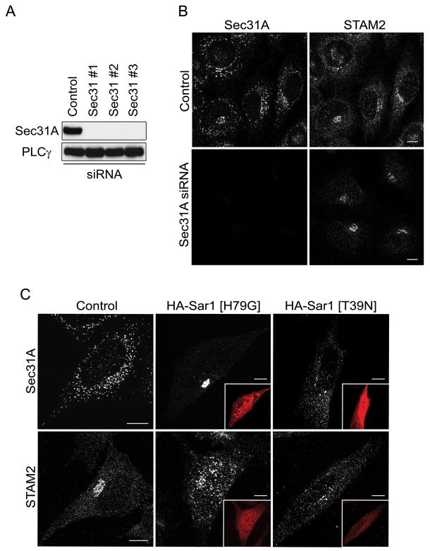
Signal-transducing adaptor molecules (STAMs) are involved in growth factor and cytokine signaling as well as receptor degradation, and they form complexes with a number of endocytic proteins, including Hrs and Eps15. In this study, we demonstrate that STAM proteins also localize prominently to early exocytic compartments and profoundly regulate Golgi morphology. Upon STAM overexpression in cells, the Golgi apparatus becomes extensively fragmented and dispersed, but when STAMs are depleted, the Golgi becomes highly condensed. Under both scenarios, vesicular stomatitis virus G protein-green fluorescent protein trafficking to the plasma membrane is markedly inhibited, and recovery of Golgi morphology after Brefeldin A treatment is substantially impaired in STAM-depleted cells. Furthermore, STAM proteins interact with coat protein II (COPII) proteins, probably at endoplasmic reticulum (ER) exit sites, and Sar1 activity is required to maintain the localization of STAMs at discrete sites. Thus, in addition to their roles in signaling and endocytosis, STAMs function prominently in ER-to-Golgi trafficking, most likely through direct interactions with the COPII complex.


Figures

Figure 1
Figure 1. STAMs interact with proteins of the early secretory pathway
A) HeLa cells were co-immunostained for endogenous STAM2 (green) and either EEA1 or GM130 (red), then visualized using confocal microscopy. Arrows in the upper panels identify an area of colocalization, and an arrowhead in a lower panel identifies an area enlarged in the insets. Bar, 10 μm. B) HeLa cell lysates were immunoprecipitated (IP) with anti-STAM1 antibodies or control IgG and immunoblotted with antibodies against endogenous STAM1, Sec31A, GGA3, GM130, AP1, or βCOP. Inputs represent 5% of the starting material. C) Lysates from cells expressing the indicated constructs were incubated with either GST or GST-STAM1 immobilized on glutathione-Sepharose beads and immunoblotted with anti-GFP antibodies. Inputs represent 5% of the starting material. D and E) HeLa cells were transfected with CFP-Sec31A (D) or Sec13-YFP (E) or else co-transfected with either construct and Myc-STAM2. Lysates were immunoprecipitated with anti-GFP antibodies and immunoblotted with antibodies against GFP or Myc-epitope. Both Sec31 and Sec13 co-precipitate with STAM2. Actin was probed to monitor specificity. An asterisk (*) in (D) denotes the IgG heavy chain. F) HeLa cells were transfected with Myc-STAM2 or else co-transfected with Myc-STAM2 and εCOP-YFP, then immunoprecipitated with anti-GFP antibodies. G) HeLa cells were singly transfected with CFP-Sec31A or co-transfected with CFP-Sec31A and HA-tagged STAM2 domains (schematic diagram on right). Cell lysates were immunoprecipitated with anti-GFP antibodies and immunoblotted with anti-GFP or anti-HA antibodies (left panels). Only the C-terminal region of STAM2 co-precipitates with Sec31 (arrowhead); asterisks (*) denote IgG heavy and light chains. HeLa cells were also singly transfected with Sec13-YFP or co-transfected with Sec13-YFP and HA-tagged STAM2 domain constructs, then immunoprecipitated with anti-GFP antibodies (middle panels). Both VHS-SH3 and C-terminal segments of STAM2 co-precipitate with Sec13 (arrowheads); asterisks (*) identify IgG heavy and light chains.
Figure 2
Figure 2. Overexpression of STAMs causes dispersion of the Golgi complex
A) Overexpression of Myc-STAM2 (left panels) in HeLa cells disperses the Golgi markers GM130 and giantin, and causes ERGIC-53 to be retained in the ER, but does not affect LAMP1 distribution (right panels). Golgi apparatus and VTCs exhibit normal, perinuclear localization patterns in untransfected cells. Arrows identify transfected cells. Bar, 10 μm. B) Quantification of Golgi fragmentation in cells overexpressing Myc-STAM1, Myc-STAM2, or empty vector (Control) (n=3; 100 cells per experiment; ±SD). *p=0.0012; **p<0.001. C) Electron microscopy images showing typical Golgi morphology in control cells (asterisk; top panel), but much smaller stacked Golgi complexes in STAM2-overexpressing cells (asterisk; bottom panel). Images are at the same magnification. Bar, 500 nm. D) Immunogold localization of GM130 in control cells (1a), with higher magnification of the boxed area in (1b). Immunogold localization of GM130 in STAM2-overexpressing cells (2a), with the boxed region enlarged in (2b). Bar, 500 nm.
Figure 3
Figure 3. Effects of STAM2 deletions on subcellular distribution and Golgi fragmentation
A) HeLa cells transfected with full-length Myc-STAM2, HA-STAM2VHS, or HA-STAM2C Term were treated with saponin to deplete cytosolic proteins and then co-stained for Myc- or HA-epitope and GM130 or, Sec61α. HeLa cells overexpressing full-length STAM2 or the C-terminal fragment display the lattice-like staining adjacent to GM130 puncta, with areas of juxtaposed staining indicated with arrows (upper panels). Colocalization of thhe Myc-STAM2 lattice-like staining with the ER protein Sec61α is shown in the middle panels. In cells expressing HA-tagged STAM2VHS, the juxtapositioned pattern with GM130 is more evident. Boxed areas are enlarged in the insets. B) Schematic diagram of STAM2 deletion constructs. Amino acid numbers are indicated. C) HeLa cells were transfected with control vector, HA-STAM2VHS, HA-STAM2VHS-SH3, HA-STAM2C Term, or full-length Myc-STAM2 (FL), co-stained with GM130 and either Myc- or HA-epitope antibodies, then assessed for Golgi fragmentation (100 cells per condition; n=3; ±SD). *p<0.01; **p<0.005; ***p<0.001.
Figure 4
Figure 4. STAM proteins colocalize with and redistribute COPII proteins
A) Full-length Myc-STAM2 and Sec13-YFP expressed in HeLa cells colocalize at multiple discrete puncta (arrows; upper panels). Sec13-YFP and HA-STAM2VHS puncta also co-localize (arrows, middle panels), sometimes juxtaposed to Golgi fragments as revealed by GM130 staining (arrowheads). The boxed area is enlarged in the lower panels. B) Overexpression of Myc-STAM2 as revealed by staining for Myc-epitope in HeLa cells results in decreased intensity of punctate staining for the COPII proteins Sec31A and Sec24C, but overexpression of STAM2 only slightly alters the Sec16L staining pattern. C) Overexpression of Myc-STAM2 has no effect on mitochondrial morphology or apoptosis as assessed with cytochrome c (Cyto c) staining (arrow indicates transfected cell). Bars, 10 μm.
Figure 5
Figure 5. STAM2 depletion results in a highly condensed Golgi apparatus
A) Lysates from HeLa cells transfected with either of two different siRNAs specific for STAM2 or else control siRNA were immunoblotted for STAM2 at 72 h post-transfection. Equal protein loading was monitored by immunoblotting for PLCγ. STAM1 levels are unchanged. B) Immunostaining of HeLa cells for STAM2 after transfection with control siRNA or STAM2 siRNA #1. Bar, 10 μm. C) HeLa cells transfected with control siRNA or STAM2 siRNA #1 were immunostained for GM130. STAM2 siRNA cells have a highly condensed Golgi apparatus and clustered ERES. Bar, 10 μm. D) Graphical representation of percentage of cells with Golgi condensation in STAM2 siRNA and control siRNA cells 72 h after transfection (n=3; 100 cells per condition; ±SD). *p <0.001. E) Graphical representation of the ratio of Golgi circumference to cell circumference in control versus STAM2 siRNA cells (n=3; 100 cells per condition). **p=0.02. F) Electron microscopy images of the Golgi complex in control cells (single asterisk; left panel) compared with that in STAM2 siRNA cells, where multiple Golgi cisternae are seen at the same magnification (asterisks; right panel). Bar, 500 nm.
Figure 6
Figure 6. Knock down and overexpression of STAM1 confirms some functional redundancy with STAM2
A) Left panel, Extracts from HeLa cells transfected with any of three siRNA oligonucleotides specific for STAM1 or else with control siRNA were immunoblotted at 72 h post-transfection. Levels of STAM2 were unaltered. Right panel, Extracts from HeLa cells transfected with control siRNA or cotransfected with siRNAs for both STAM1 and STAM2 (STAM DKD) were immunoblotted with the indicated antibodies. Equal protein loading was monitored using PLCγ. B) HeLa cells were transfected with STAM1 siRNA or co-transfected with siRNAs for STAM1 and STAM2 (STAM double knock down [DKD]) or else with control siRNA. Percentages of cells with Golgi condensation (≤5 μm diameter in all dimensions) in STAM1 siRNA, STAM DKD, and control siRNA groups (n=3; 100 cells per experiment) are presented graphically (±SD). *p=0.0042; **p<0.0001. C) HeLa cells transfected with control, STAM1, or STAM DKD siRNAs were immunostained with anti-GM130 antibodies. As observed for STAM2 siRNA knock down cells, STAM1 siRNA cells also have a highly condensed Golgi apparatus. Bar, 10 μm. D) Re-transfection of HeLa cells with Myc-STAM1 after transfection with STAM2 siRNA suppressed the condensed Golgi phenotype (arrow). Bar, 10 μm.
Figure 7
Figure 7. Localization of STAM2 to ERES in BFA-treated cells and delayed recovery of Golgi morphology in STAM2 siRNA cells following BFA
A) HeLa cells were treated with BFA and co-stained for STAM2 (green) and GM130 or Sec31A (red). STAM2 puncta are adjacent to GM130 puncta, and STAM2 puncta colocalize with Sec31A puncta. B) HeLa cells transfected with control or STAM2 siRNAs were treated with BFA and assessed after wash-out. Most control cells (upper panels) display normal Golgi morphology as assessed by GM130 staining after 1 h of recovery, whereas most STAM2 siRNA-treated cells (lower panels) have not recovered even after 3 h. Bar, 10 μm. C) Graphical presentation of the percentage of cells with Golgi reconstitution (lacking tubular Golgi) after transfection with STAM2 siRNA and control siRNA (n=3; 100 cells per experiment; ±SD).
Figure 8
Figure 8. VSVG trafficking is impaired in cells lacking or overexpressing STAM2
A) HeLa cells were transfected with control siRNA (top panels) or STAM2 siRNA (bottom panels), and then re-transfected with ts045 VSVG-GFP. VSVG trafficked to the plasma membrane as early as 60 min after moving to a permissive temperature in control cells, while trafficking to plasma membrane was substantially reduced in the STAM2 siRNA cells. B) HeLa cells were singly transfected with VSVG-GFP (top panels) or else co-transfected with Myc-STAM2 (bottom panels; Myc staining not shown), then assessed at different time points after moving to a temperature permissive for VSVG trafficking. VSVG is present at the plasma membrane in control cells as soon as 90 min after the temperature change, whereas it is retained within the fragmented Golgi of STAM2-overexpressing cells even at 180 min. C) VSVG and GM130 are present in the same fragmented Golgi compartments in HeLa cells overexpressing STAM2 180 min after temperature change. The enlarged image in the inset shows VSVG staining (green) adjacent to GM130 puncta (red). Bar, 10 μm.
Figure 9
Figure 9. STAM2 recruitment to the early secretory pathway is dependent on Sar1 activity, but not on Sec31
A) Lysates from HeLa cells transfected with any of three different Sec31A siRNAs or else control siRNA were immunoblotted for Sec31A 72 h post-transfection. Equal protein loading was monitored using PLCγ. B) HeLa cells were co-stained with antibodies against STAM2 and Sec31A after transfection with control or Sec31A siRNAs; there is no change in STAM2 distribution upon Sec31A depletion. C) Control HeLa cells were stained for STAM2 or Sec31A (left panels). Cells transfected with HA-Sar1 [H79G] (HA-epitope staining in insets) show changes in distribution patterns for both proteins, with STAM2 localizing to large punctate structures and Sec31A labeling becoming more condensed (middle panels). Cells expressing HA-Sar1 [T39N] (HA-epitope staining in insets) show decreased staining intensity of puncta and dispersal of immunoreactivity for both STAM2 and Sec31A (right panels).

References

    1. Endo K, Takeshita T, Kasai H, Sasaki Y, Tanaka N, Asao H, Kikuchi K, Yamada M, Chenb M, O’Shea JJ, Sugamura K. STAM2, a new member of the STAM family, binding to the Janus kinases. FEBS Lett. 2000;477:55–61. - PubMed
    1. Pandey A, Fernandez MM, Steen H, Blagoev B, Nielsen MM, Roche S, Mann M, Lodish HF. Identification of a novel immunoreceptor tyrosine-based activation motif-containing molecule, STAM2, by mass spectrometry and its involvement in growth factor and cytokine receptor signaling pathways. J Biol Chem. 2000;275:38633–38639. - PubMed
    1. Takeshita T, Arita T, Asao H, Tanaka N, Higuchi M, Kuroda H, Kaneko K, Munakata H, Endo Y, Fujita T, Sugamura K. Cloning of a novel signal-transducing adaptor molecule containing an SH3 domain and ITAM. Biochem Biophys Res Commun. 1996;225:1035–1039. - PubMed
    1. Takeshita T, Arita T, Higuchi M, Asao H, Endo K, Kuroda H, Tanaka N, Murata K, Ishii N, Sugamura K. STAM, signal transducing adaptor molecule, is associated with Janus kinases and involved in signaling for cell growth and c-myc induction. Immunity. 1997;6:449–457. - PubMed
    1. Lohi O, Lehto V-P. STAM/EAST/Hbp adapter proteins--integrators of signalling pathways. FEBS Lett. 2001;508:287–290. - PubMed

Publication types

MeSH terms

Substances

LinkOut - more resources