. 2009 Jan 30;284(5):3086-3095.
doi: 10.1074/jbc.M803602200. Epub 2008 Dec 3.

CCAAT enhancer-binding protein alpha is a molecular target of 1,25-dihydroxyvitamin D3 in MCF-7 breast cancer cells

Affiliations

CCAAT enhancer-binding protein alpha is a molecular target of 1,25-dihydroxyvitamin D3 in MCF-7 breast cancer cells

Puneet Dhawan et al. J Biol Chem. .

Erratum in

  • J Biol Chem. 2009 Mar 20;284(12):8208. Weider, Robert [corrected to Wieder, Robert]

Abstract

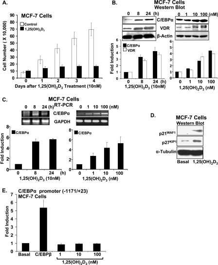
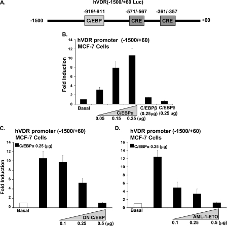
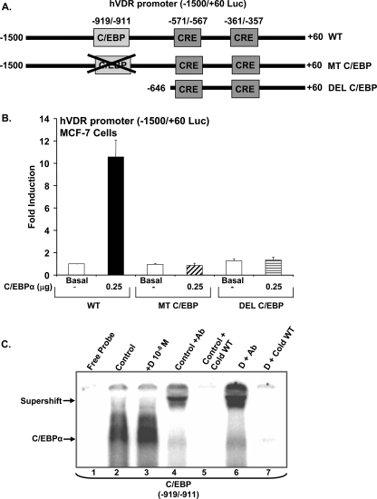
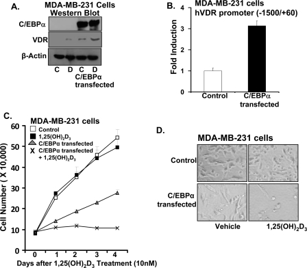
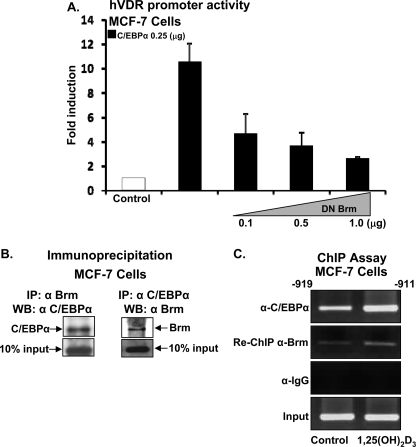
Numerous studies have shown that the active form of vitamin D, 1,25(OH)(2)D(3), can exert growth inhibitory effects on human breast cancer cells and mammary tumor growth. However, the molecular mechanisms remain to be fully delineated. This study demonstrates for the first time that CCAAT enhancer-binding protein alpha (C/EBPalpha), a member of the C/EBP family of transcription factors, is induced by 1,25(OH)(2)D(3) and is a potent enhancer of VDR transcription in MCF-7 breast cancer cells. 1,25(OH)(2)D(3) was found to induce C/EBPalpha as well as VDR expression in MCF-7 cells. C/EBPalpha was not detected in MDA-MB-231 cells that are poorly responsive to 1,25(OH)(2)D(3). Antiproliferative effects of 1,25(OH)(2)D(3) and induction of VDR were observed in MDA-MB-231 cells transfected with C/EBPalpha, and knockdown of C/EBPalpha suppressed VDR and antiproliferative effects of 1,25(OH)(2)D(3) in MCF-7 cells. Transfection of C/EBPalpha in MCF-7 cells resulted in a dose-dependent enhancement of hVDR transcription. Our studies show that C/EBPalpha can bind to Brahma (Brm), an ATPase that is a component of the SWI/SNF complex, and cooperate with Brm in the regulation of hVDR transcription in MCF-7 cells. Because the levels of VDR in MCF-7 breast cancer cells correlate with the antiproliferative effects of 1,25(OH)(2)D(3) and because C/EBPalpha has been suggested as a potential tumor suppressor in breast cancer, these findings provide important mechanisms whereby 1,25(OH)(2)D(3) may act to inhibit growth of breast cancer cells. These findings also identify C/EBPalpha as a 1,25(OH)(2)D(3) target in breast cancer cells and provide evidence for C/EBPalpha as a candidate for breast cancer treatment.


Figures

FIGURE 1.
FIGURE 1.
C/EBPα is induced by 1,25(OH)2d3 in MCF-7 breast cancer cells. A, proliferation of MCF-7 cells is significantly decreased in the presence of 1,25(OH)2d3 (10 nm) at all times examined (1–4 days, p < 0.05 compared with vehicle). Data represent the mean ± S.E. of three independent experiments. B, left panel top, representative Western blot. Western blot was performed using nuclear extracts from MCF-7 cells treated with vehicle or 1,25(OH)2d3 (10 nm for 8–24 h) and probed with C/EBPα, VDR, and β-actin antibodies. Left panel bottom, graphic representation of densitometric scans of Western blots. Data represent the mean ± S.E. of three independent experiments. C/EBPα (42 kDa) and VDR (48 kDa) are significantly induced by 1,25(OH)2d3 at 8 and 24 h (p < 0.05 compared with 0 time). Right panel top, 1,25(OH)2d3 dose response; representative Western blot. Nuclear extracts from MCF-7 cells treated with vehicle or increasing concentrations of 1,25(OH)2d3 (1–100 nm) for 24 h were used. Right panel bottom, graphic representation of densitometric scans of Western blots. C/EBPα and VDR are significantly induced at 10 and 100 nm 1,25(OH)2d3 (p < 0.05 compared with vehicle control). Data represent the mean ± S.E. of three independent experiments. C, top panels: representative RT-PCR analyses of mRNA from MCF-7 cells for C/EBPα. Cells were treated with 1,25(OH)2d3 as in Fig. 1B. Bottom panels: quantitation of C/EBPα mRNA expression. Data represent the mean ± S.E. of three independent experiments. C/EBPα mRNA is significantly induced by 1,25(OH)2d3 at 8 and 24 h (left panel, p < 0.05 compared with 0 time). Right panel: C/EBPα mRNA is significantly induced at 1, 10, and 100 nm 1,25(OH)2d3 (p < 0.05 compared with vehicle control). D, Western blot analysis of MCF-7 cells treated with vehicle or 1,25(OH)2d3 (10 nm) for 24 h using p21, p27, and α-tubulin antibodies. The Western blot is representative of results obtained from at least two additional experiments. E, 1,25(OH)2d3 does not affect the activity of the C/EBPα promoter. MCF-7 cells transfected with the C/EBPα promoter (-1171/+23) (65) were co-transfected with the C/EBPβ expression plasmid (0.25 μg) or treated with 1,25(OH)2d3 (1–100 nm). The data represent the mean ± S.E. of three separate experiments. The induction of C/EBPα promoter activity by C/EBPβ (as previously reported (65)) is significant at p < 0.05 compared with cells transfected with vector alone. No significant change in promoter activity was observed after 1,25(OH)2d3 treatment (p > 0.5).
FIGURE 2.
FIGURE 2.
Enhancement of hVDR promoter transcription by C/EBPα in MCF-7 breast cancer cells. A, schematic of the luciferase construct of the human VDR promoter -1500/+60, including a putative C/EBP motif and two putative CRE sites. B, MCF-7 cells were co-transfected with hVDR promoter and increasing concentrations of C/EBPα (0.05 μg to 0.25 μg) or 0.25 μg of C/EBPβ or C/EBPδ. C/EBPα (0.05, 0.15, and 0.25 μg) results in a significant induction in hVDR promoter activity (p < 0.05 compared with basal). C and D, suppression of hVDR promoter transcription using DN C/EBP or AML-1/ETO. MCF-7 cells were co-transfected hVDR promoter, 0.25 μg of C/EBPα and increasing concentrations of DN C/EBP or of AML-1/ETO, a C/EBPα inhibitor. Empty vectors were used to keep the total DNA concentrations the same. In MCF-7 cells there was no effect of DN CEBP or AML-1/ETO at the concentrations used on basal levels of hVDR transcription. pRL-TF-Renilla luciferase was co-transfected as an internal control. Results of three or more separate experiments are presented (mean ± S.E.). Co-transfection with 0.25 μg or 0.5 μg of DN C/EBP or 0.1, 0.25, or 0.5 μg of AML-1/ETO resulted in a significant decrease in C/EBPα-induced VDR transcription (p < 0.05 compared with cells transfected with C/EBPα (0.25 μg) alone).
FIGURE 3.
FIGURE 3.
Detection of C/EBPα activation domain in the hVDR promoter. A, schematic of luciferase constructs of the hVDR promoter and mutation and deletions of the C/EBP site. hVDR promoter construct (-1500/+60) (WT); hVDR (-1500/+60) promoter with the putative C/EBP site mutated (MT C/EBP) and hVDR promoter -646/+60 with C/EBP putative site deleted (DEL C/EBP) are shown. B, luciferase assay determined in extracts of MCF-7 cells transfected using luciferase constructs of hVDR promoter shown in A with co-transfection of C/EBPα expression vector (0.25 μg) or vector alone (basal). Luciferase activity is represented as -fold induction over the control (mean ± S.E.; three to six observations per group). pRL-TK-Renilla luciferase was co-transfected as an internal control. C, identification of C/EBPα binding motif in the hVDR promoter by electrophoretic mobility shift assay. The 32P-labeled oligonucleotide probes containing wild-type sequence were incubated with 5 μg of nuclear protein from vehicle (lane 2) or 1,25(OH)2d3-treated (lane 3) MCF-7 cells transfected with the C/EBPα expression vector. The wild-type probe was incubated with C/EBPα antibody in the presence of 5 μg of vehicle treated (lane 4) or 1,25(OH)2d3-treated (lane 6) nuclear protein. The wild-type probe in presence of vehicle treated (lane 5) or 1,25(OH)2d3-treated (lane 7) nuclear protein was also incubated with a 100-fold molar excess of wild-type (WT) cold competitor oligonucleotide. Gel mobility shift data are representative of at least three experiments.
FIGURE 4.
FIGURE 4.
Inhibition of C/EBPα expression using siRNA reduces the anti-proliferative effect of 1,25(OH)2d3 in MCF-7 cells. A, sequences of C/EBPα siRNA and scrambled siRNA used for transfection of MCF-7 cells. B, Western blot analysis of C/EBPα and VDR in control, C/EBPα siRNA-transfected, and scrambled siRNA-transfected MCF-7 cells. Two additional experiments yielded similar results. C, graphic representation of cell number of control, C/EBPα siRNA-transfected, and scrambled siRNA-transfected MCF-7 cells treated with vehicle or 10 nm 1,25(OH)2d3 as described under “Experimental Procedures.” Results represent the mean ± S.E. of three separate experiments.
FIGURE 5.
FIGURE 5.
Effect of C/EBPα expression in MDA-MB-231 cells. A, Western blot analysis. MDA-MB-231 cells were transfected with green fluorescent protein expression vector (with G418 resistance) and C/EBPα. Cells were selected on increasing concentration of G418 (200–600 μg/ml) for 2 weeks. Western blot was performed using nuclear extracts from control or C/EBPα-transfected cells treated with vehicle or 1,25(OH)2d3 (10 nm) for 24 h. Detection was by immunoblotting using C/EBPα, VDR, and β-actin antibodies. Two additional experiments yielded similar results. B, control or C/EBPα-transfected MDA-MB-231 cells were transfected with hVDR promoter (0.5 μg). C/EBPα-transfected cells show a significant increase in hVDR promoter activity compared with control, vector-transfected cells (p < 0.05 compared with control). C, proliferation of MDA-MB-231 cells in response to 1,25(OH)2d3 in the presence of absence of C/EBPα. Cells were transfected with vector or C/EBPα as described above and treated with vehicle or 1,25(OH)2d3 (10 nm). Differences in proliferation between MDA-MB-231 cells treated with vehicle or 1,25(OH)2d3 were not observed. C/EBPα expression resulted in a significant decrease in MDA-MB-231 cell proliferation (1–4 days; p < 0.05 compared with control vector-transfected cells). 1,25(OH)2d3 significantly enhanced the inhibition of growth in C/EBPα-transfected MDA-MB-231 cells (1–4 days; p < 0.05 compared with C/EBPα-transfected cells). D, light microscopic image of MDA-MB-231 cells in the presence or absence of C/EBPα treated for 4 days with vehicle or 1,25(OH)2d3 (10 nm) (magnification, 200×).
FIGURE 6.
FIGURE 6.
Functional cooperation between C/EBPα and SWI/SNF complex. A, MCF-7 cells were co-transfected with the hVDR promoter luciferase construct -1500/+60 and C/EBPα expression plasmid (0.25 μg) in the presence or absence of increasing concentrations of dominant negative BRM. Co-transfection with DN Brm resulted in a significant decrease in VDR promoter activity at all concentrations used (p < 0.05 compared with cells transfected with C/EBPα (0.25 μg) alone). B, co-immunoprecipitation of C/EBPα and Brm. Left panel: MCF-7 cells were lysed and immunoprecipitated with BRM antibody and Western blots were performed using C/EBPα antibody. Right panel: cells were lysed and immunoprecipitated with C/EBPα antibody, and Brm was detected in the immunoprecipitate using Brm antibody. These findings are representative of three separate experiments. Enhanced interaction was not observed when immunoprecipitation was done in the presence of 10 nm 1,25(OH)2d3 (not shown). C, C/EBPα and Brm are recruited to the VDR promoter in intact cells. MCF-7 cells were treated with vehicle or 1,25(OH)2d3 (10 nm) for 24 h, cells were cross-linked, and cell lysates were subjected to immunoprecipitation first with C/EBPα antibody and then with Brm antibody. DNA was isolated and PCR, using specific primers designed against the C/EBP site on the hVDR promoter, was performed. PCR was carried out in the linear range of DNA amplification. Immunoprecipitation with IgG was used as control (IgG panel). These experiments are representative of three separate experiments performed under the similar condition.

References

    1. Christakos, S., Dhawan, P., Liu, Y., Peng, X., and Porta, A. (2003) J. Cell Biochem. 88 695-705 - PubMed
    1. Jacobson, E. A., James, K. A., Newmark, H. L., and Carroll, K. K. (1989) Cancer Res. 49 6300-6303 - PubMed
    1. Colston, K. W., Chander, S. K., Mackay, A. G., and Coombes, R. C. (1992) Biochem. Pharmacol. 44 693-702 - PubMed
    1. Colston, K. W., Mackay, A. G., James, S. Y., Binderup, L., Chander, S., and Coombes, R. C. (1992) Biochem. Pharmacol. 44 2273-2280 - PubMed
    1. Anzano, M. A., Smith, J. M., Uskokovic, M. R., Peer, C. W., Mullen, L. T., Letterio, J. J., Welsh, M. C., Shrader, M. W., Logsdon, D. L., and Driver, C. L., Brown, C. C., Roberts A. B., and Sporn, M. B. (1994) Cancer Res. 54 1653-1656 - PubMed

Publication types

MeSH terms