

Regulation of cadherin trafficking
- PMID: 19055694
- PMCID: PMC2905039
- DOI: 10.1111/j.1600-0854.2008.00862.x
Regulation of cadherin trafficking
Abstract
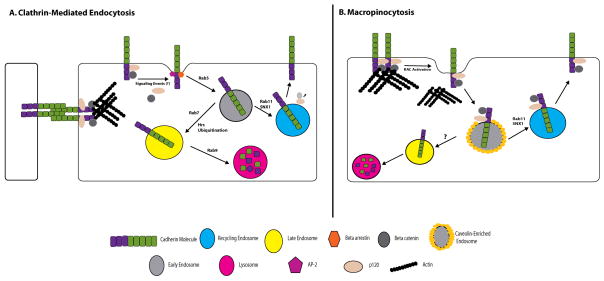
Cadherins are a large family of cell-cell adhesion molecules that tether cytoskeletal networks of actin and intermediate filaments to the plasma membrane. This function of cadherins promotes tissue organization and integrity, as demonstrated by numerous disease states that are characterized by the loss of cadherin-based adhesion. However, plasticity in cell adhesion is often required in cellular processes such as tissue patterning during development and epithelial migration during wound healing. Recent work has revealed a pivotal role for various membrane trafficking pathways in regulating cellular transitions between quiescent adhesive states and more dynamic phenotypes. The regulation of cadherins by membrane trafficking is emerging as a key player in this balancing act, and studies are beginning to reveal how this process goes awry in the context of disease. This review summarizes the current understanding of how cadherins are routed and how the interface between cadherins and membrane trafficking pathways regulates cell surface adhesive potential. Particular emphasis is placed on the regulation of cadherin trafficking by catenins and the interplay between growth factor signaling pathways and cadherin endocytosis.
Figures
References
-
- Wheelock MJ, Johnson KR. Cadherins as modulators of cellular phenotype. Annual review of cell and developmental biology. 2003;19:207–235. - PubMed
-
- Holthofer B, Windoffer R, Troyanovsky S, Leube RE. Structure and function of desmosomes. International review of cytology. 2007;264:65–163. - PubMed
-
- Green KJ, Gaudry CA. Are desmosomes more than tethers for intermediate filaments? Nature reviews. 2000;1(3):208–216. - PubMed
-
- Emery G, Knoblich JA. Endosome dynamics during development. Current opinion in cell biology. 2006;18(4):407–415. - PubMed
-
- Mattey DL, Garrod DR. Splitting and internalization of the desmosomes of cultured kidney epithelial cells by reduction in calcium concentration. Journal of cell science. 1986;85:113–124. - PubMed
Publication types
MeSH terms
Substances
Grants and funding
LinkOut - more resources
Full Text Sources
