. 2008 Dec 3:6:77.
doi: 10.1186/1479-5876-6-77.

A highly invasive human glioblastoma pre-clinical model for testing therapeutics

Affiliations

A highly invasive human glioblastoma pre-clinical model for testing therapeutics

Qian Xie et al. J Transl Med. .

Abstract

Animal models greatly facilitate understanding of cancer and importantly, serve pre-clinically for evaluating potential anti-cancer therapies. We developed an invasive orthotopic human glioblastoma multiforme (GBM) mouse model that enables real-time tumor ultrasound imaging and pre-clinical evaluation of anti-neoplastic drugs such as 17-(allylamino)-17-demethoxy geldanamycin (17AAG). Clinically, GBM metastasis rarely happen, but unexpectedly most human GBM tumor cell lines intrinsically possess metastatic potential. We used an experimental lung metastasis assay (ELM) to enrich for metastatic cells and three of four commonly used GBM lines were highly metastatic after repeated ELM selection (M2). These GBM-M2 lines grew more aggressively orthotopically and all showed dramatic multifold increases in IL6, IL8, MCP-1 and GM-CSF expression, cytokines and factors that are associated with GBM and poor prognosis. DBM2 cells, which were derived from the DBTRG-05MG cell line were used to test the efficacy of 17AAG for treatment of intracranial tumors. The DMB2 orthotopic xenografts form highly invasive tumors with areas of central necrosis, vascular hyperplasia and intracranial dissemination. In addition, the orthotopic tumors caused osteolysis and the skull opening correlated to the tumor size, permitting the use of real-time ultrasound imaging to evaluate antitumor drug activity. We show that 17AAG significantly inhibits DBM2 tumor growth with significant drug responses in subcutaneous, lung and orthotopic tumor locations. This model has multiple unique features for investigating the pathobiology of intracranial tumor growth and for monitoring systemic and intracranial responses to antitumor agents.


Figures

Figure 1
Figure 1
In an experimental metastasis model, DBM2 cells produce tumors in various tissues. (A) Clonal selection through experimental metastasis. The DBTRG-05MG cells were injected into the tail vein of athymic nude mice. Mice were sacrificed either when they became moribund (~12 weeks) or after 8 weeks. At necropsy, lung lesions were transplanted into nude mice subcutaneously. From these tumors, cells were harvested and injected into nude mice via tail vein. After the second cycle (M2) cells were expanded ex-vivo in culture. (B) DBTRG-05MG or DBM2 cells were injected via the tail vein into nude mice. After eight weeks mice inoculated with DBTRG-05MG cells had only a few pulmonary tumors (a, b). By contrast, lungs from mice bearing DBM2 cells were almost fully replaced with tumors (c, d), and metastatic foci were found in skeletal muscle (e), diaphragm (f), lymph nodes adjacent to the spinal cord (g) and in the chest cavity (h). H&E staining of formalin fixed sections from lungs of DBTRG-05MG cells (i) or DBM2 cells (j) eight weeks after tail vein injection. Invasion of DBM2 tumors into skeletal muscle (left 2 arrows) induces bone resorption (right arrow) (k) and replaces nearly the entire lymph node (arrow) (l, insert at low magnification).
Figure 2
Figure 2
Elevated cytokines and growth factors in GBM-M2 cells. Identification of cytokines and growth factors in common in the 24 hr conditioned medium for all three GBM-M2 tumor lines and the fold increases in their expression compared to the parental GBM cells. Heat map shows fold differences based upon the of expression ratios of 89 cytokines and proteins between parental and GBM-M2 lines determined as described in the materials and methods section. The fold change in protein expression level is indicated by color. GM-CSF, IL-6, IL-8 and BDNF were found highly elevated in all three GBM-M2 lines (fold changes are summarized in the supplementary table [Additional File 4]).
Figure 3
Figure 3
Invasive growth and GBM properties of orthotopic DBM2 intracranial tumors. (A) Orthotopic DBM2 tumors exhibit extensive infiltration into the mouse brain parenchyma (a, b). The arrows point to areas of cranial erosion. (c) Higher magnification of DBM2 tumor demonstrating extensive infiltration into the brain parenchyma. Compared to DBM2, U251 tumors form a sharper cranial margin (d, e) and are less invasive (f). (B) Met (a, b) and uPAR (c, d) expression in invasive DMB2 orthotopic tumors. (C) H&E staining of formalin fixed DBM2 tumors shows central necrosis with the crowding of cancer cells lining the necrotic area (a, b arrows). Vascular invasion of DBM2 tumors along the perivascular space (arrow) and in vessels in the surrounding brain (c) with tumor-thrombus formation (d). Higher magnification showing a glomeruloid body-like structure (d, insert). CD31 staining highlights vascular proliferation (e). Enlargement of (e) showing glomeruloid body-like structure with multiple layers of endothelial cells is stained by CD31 antibody (f).
Figure 4
Figure 4
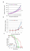
17 AAG inhibition of DBM2 tumor growth. (A) 17AAG at 60 mg/kg-d inhibits DBM2 subcutaneous tumor growth. DBM2 cells were inoculated into the flanks of nude mice at 5 × 10⁵ cells in 100 ul PBS. After 2 weeks, mice with size-matched tumors (100 – 200 mm3) were assigned into control and treatment (60 mg/kg-d) groups (n = 19) and treatment started. Error bar represents for standard error. (B) 17AAG at 60 mg/kg-d inhibits DBM2 orthotopic tumor growth. DBM2 cells were inoculated intracranially into nude mice at 5 × 10⁵ cells in 5 ul PBS. The tumor growth was monitored by Ultrasound. After 2 weeks, size-matched tumors were grouped into control and treatment groups (n = 10). Fold change of tumor volume = Weekly measured tumor size/Initial tumor size upon grouping. (C) The survival time of nude mice bearing orthotopic DBM2 tumor xenografts treated with 17AAG. DBM2 cells were inoculated intracranially of nude mice at 5 × 10⁵ cells in 5 ul PBS. After 3 weeks, size-matched tumors were grouped into control (n = 6) and 2 treatment groups (20 mg/kg, 60 mg/kg, n = 8). The arrow points to the day treatment started after orthotopic tumor inoculation. Treatment was administered until individual mice became moribund according to IACUC guild-line and survival time was recorded.

References

    1. Berens ME, Giese A. "...those left behind." Biology and oncology of invasive glioma cells. Neoplasia. 1999;1:208–219. doi: 10.1038/sj.neo.7900034. - DOI - PMC - PubMed
    1. Camphausen K, Purow B, Sproull M, Scott T, Ozawa T, Deen DF, Tofilon PJ. Influence of in vivo growth on human glioma cell line gene expression: convergent profiles under orthotopic conditions. Proc Natl Acad Sci USA. 2005;102:8287–8292. doi: 10.1073/pnas.0502887102. - DOI - PMC - PubMed
    1. Assanah M, Lochhead R, Ogden A, Bruce J, Goldman J, Canoll P. Glial progenitors in adult white matter are driven to form malignant gliomas by platelet-derived growth factor-expressing retroviruses. J Neurosci. 2006;26:6781–6790. doi: 10.1523/JNEUROSCI.0514-06.2006. - DOI - PMC - PubMed
    1. Lee J, Kotliarova S, Kotliarov Y, Li A, Su Q, Donin NM, Pastorino S, Purow BW, Christopher N, Zhang W, et al. Tumor stem cells derived from glioblastomas cultured in bFGF and EGF more closely mirror the phenotype and genotype of primary tumors than do serum-cultured cell lines. Cancer Cell. 2006;9:391–403. doi: 10.1016/j.ccr.2006.03.030. - DOI - PubMed
    1. Bao S, Wu Q, McLendon RE, Hao Y, Shi Q, Hjelmeland AB, Dewhirst MW, Bigner DD, Rich JN. Glioma stem cells promote radioresistance by preferential activation of the DNA damage response. Nature. 2006;444:756–760. doi: 10.1038/nature05236. - DOI - PubMed

Publication types

MeSH terms

LinkOut - more resources