Review
. 2009 Mar 15;128(1-3):147-70.
doi: 10.1016/j.vetimm.2008.10.321. Epub 2008 Nov 1.

The piglet as a model for B cell and immune system development

Affiliations
Review

The piglet as a model for B cell and immune system development

J E Butler et al. Vet Immunol Immunopathol. .

Abstract

The ability to identify factors responsible for disease in all species depends on the ability to separate those factors which are environmental from those that are intrinsic. This is particularly important for studies on the development of the adaptive immune response of neonates. Studies on laboratory rodents or primates have been ambiguous because neither the effect of environmental nor maternal factors on the newborn can be controlled in mammals that: (i) transmit potential maternal immunoregulatory factors in utero and (ii) are altricial and cannot be reared after birth without their mothers. Employing the newborn piglet model can address each of these concerns. However, it comes at the price of having first to characterize the immune system of swine and its development. This review focuses on the porcine B cell system, especially on the methods used for its characterization in fetal studies and neonatal piglets. Understanding these procedures is important in the interpretation of the data obtained. Studies on neonatal piglets have (a) provided valuable information on the development of the adaptive immune system, (b) lead to important advances in evolutionary biology, (c) aided our understanding of passive immunity and (d) provided opportunities to use swine to address specific issues in veterinary and biomedical research and immunotherapy. This review summarizes the history of the development of the piglet as a model for antibody repertoire development, thus providing a framework to guide future investigators.
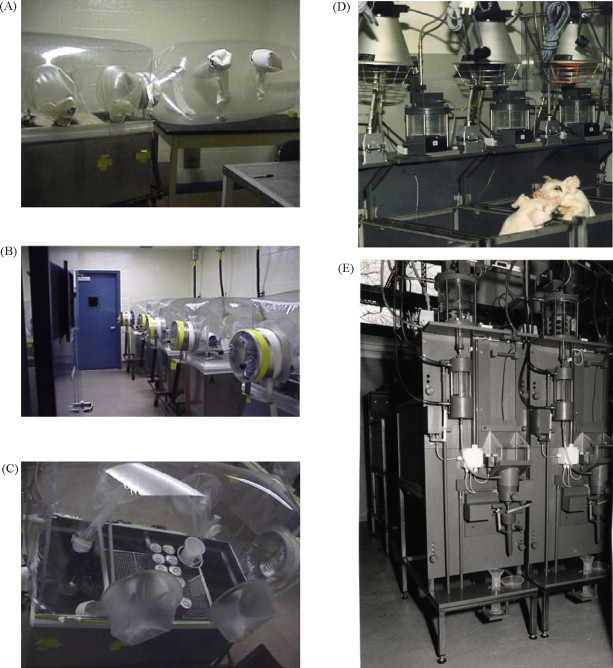

Figures

Fig. 1
Fig. 1
Transfer of immunity from mother to young among common placental mammals. Group I and Group III mammals represent the extremes in which receptor-mediated transport of IgG occurs via the placenta (Grp I) versus the mammary gland (Grp III). This difference is reflected in the Ig constituency of the colostrum; font size depicts relative Ig concentrations in colostrum. Uptake by the suckling neonate differs among mammals. In rodents this is mediated by FcRn which transports IgG while among Grp III mammals it is receptor independent (Butler, 1974).
Fig. 2
Fig. 2
The critical window of immunological development. The period between late gestation and weaning is depicted as a window in which natural and passive antibodies provide protection to the neonate and when colonization drives the development of adaptive immunity including oral tolerance. The dotted line indicates the period in which natural antibodies are considered important. Occurring in an orderly and regulated fashion, the events during this period create a state of immune homeostasis by the time of weaning (Butler et al., 2006, Butler and Sinkora, 2007).
Fig. 3
Fig. 3
(Top) Diversity in organs and pathways involved in B cell repertoire development among selected vertebrates. Left: mouse and human. Right: chicken, rabbits and perhaps hoofed mammals. (Bottom) The relative importance of mechanisms used by different vertebrates in generating their heavy chain Ab repertoire. SHM = somatic hypermutation; Comb. or C = combinatorial diversity; Junct. or junctional = junctional diversity.
Fig. 4
Fig. 4
Class switch recombination to IgG occurs during phase 2 of gestation and favors expression of IgG1 throughout fetal life except in the IPP and MLN in late gestation when IgG3 is preferentially expressed in theses organs. IgG3 expression is temporally linked to the time when the IPP is believed to function as a primary lymphoid organ (Butler and Wertz, 2006).
Fig. 5
Fig. 5
The comparative amino acid sequence of VpreB from swine, mouse and human.
Fig. 6
Fig. 6
Gnotobiotic and autosow facilities. (A) Gnotobiotic isolator at SDSU with the surgery bubble attached to isolator entry port. (B) Gnotobiotic isolators shown from port end. The exterior port cap is taped in place to prevent loss due to interior pressure when the interior port cap is removed. The interior port cap is visible through the isolator canopy in the first isolator. (C) View from the top of a gnotobiotic the isolator through isolator canopy. A stainless steel wire mesh rack is fixed to the top of the isolator pen dividers and is used for storage of milk replacer and other supplies. The can in the port contains potassium permanganate used as a catalyst to boil and vaporize the formaldehyde sterilizing solution. The yellow band around the lip of the isolator is a motorcycle clamp used to prevent internal air pressure from pealing tape away from isolator. (D) The autosow facilities at the Institute of Animal Husbandry, Mariensee, Germany. Each animal is housed separately so that an accurate measurement of the amount of milk or milk replacer consumed can be determined. Feeding time is computer controlled. The mixing and warming chamber for the milk is visible just below the thermostatically controlled heat lamps. (E) Back side of the autosow feeding device showing the system used to measure the amount of milk consumed and the chambers containing the wash and rinse fluids of the automatic feeder washing system.
Fig. 7
Fig. 7
The IgM and IgG Ab response to naked TD (FLU-KLH) and TI-2 (TNP-Ficoll) antigens by germfree piglets (GF) and piglets monoassociated with benign Escherichia coli (colonized). Naked Antigen = an antigen given without natural or artificial adjuvants.

References

    1. Ackermann M. Respiratory tract. In: Pond J.E., Mersmann H.J., editors. Biology of the Domestic Pig. Cornell University Press; New York: 2001. pp. 502–532.
    1. Allen W.D., Porter P. The demonstration of immunoglobulins in porcine intestinal tissue by immunofluorescence with observations on the effect of fixation. Immunology. 1970;18:799–806. - PMC - PubMed
    1. Allen W.D., Porter P. The relative distribution of IgM and IgG cells in intestinal ucosa and lymphoid tissue of the young unweaned pig and their significance in ontogenesis of secretory immunity. Immunology. 1973;24:493–501. - PMC - PubMed
    1. Allen W.D., Porter P. Localization of immunofluorescence of secretory component and IgA in intestinal mucosa of the young pig. Immunology. 1973;24:365–374. - PMC - PubMed
    1. Atkins A.M., Schofield G.C., Reeders T. Studies on the structure and distribution of immunoglobulin A containing cells in the gut of the pig. J. Anat. 1971;109:385–395. - PMC - PubMed