. 2009 Feb 1;326(1):177-89.
doi: 10.1016/j.ydbio.2008.11.009. Epub 2008 Nov 20.

Phosphorylation of Gli2 by protein kinase A is required for Gli2 processing and degradation and the Sonic Hedgehog-regulated mouse development

Affiliations

Phosphorylation of Gli2 by protein kinase A is required for Gli2 processing and degradation and the Sonic Hedgehog-regulated mouse development

Yong Pan et al. Dev Biol. .

Abstract

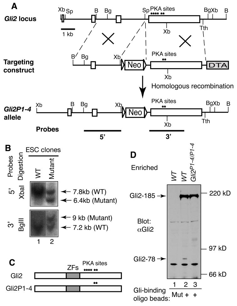
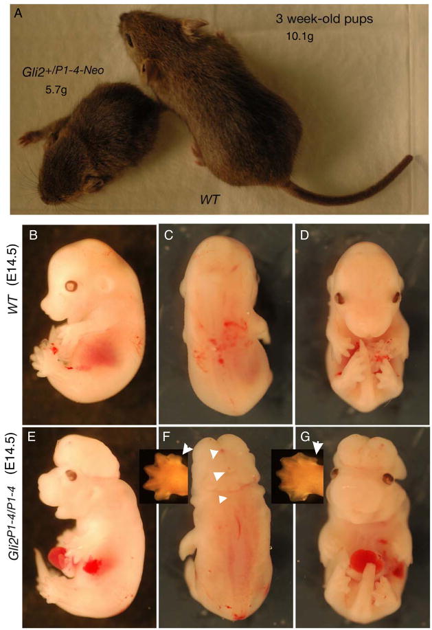
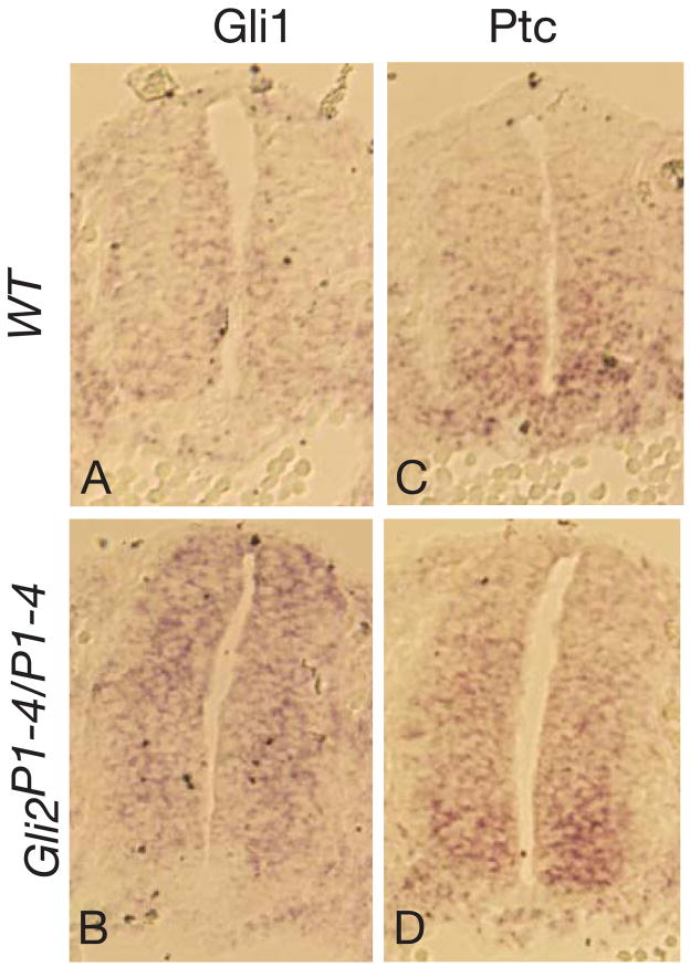
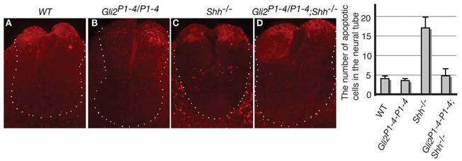
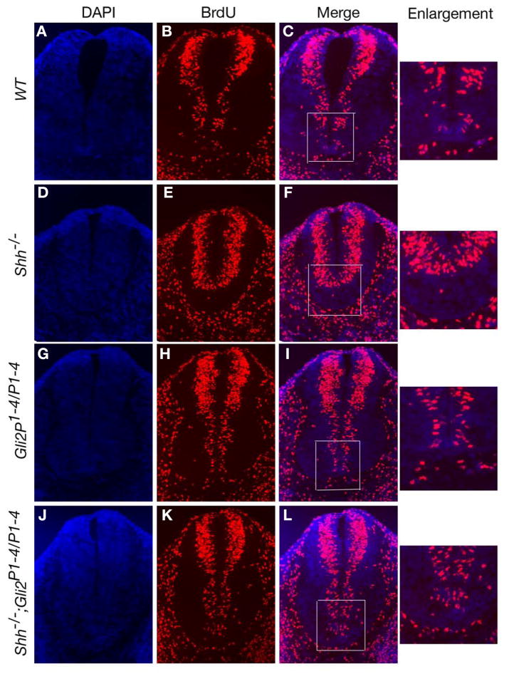
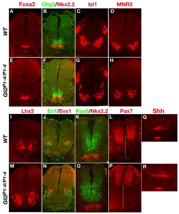
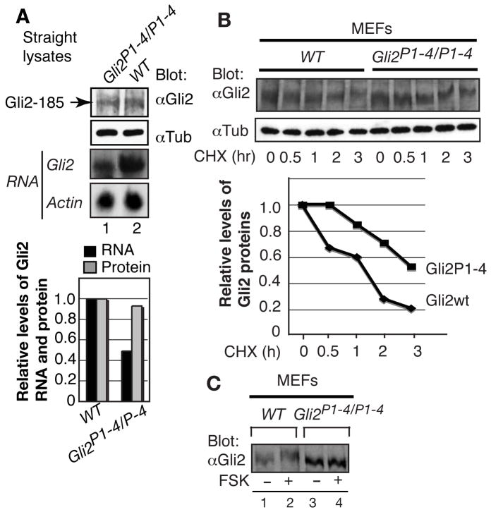
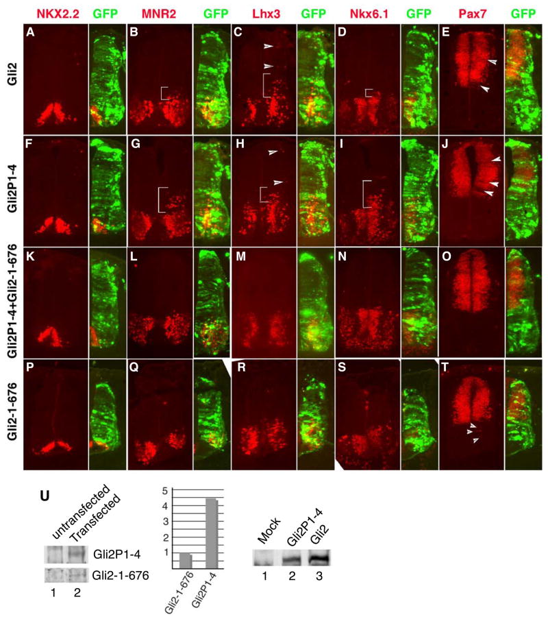
In mice, Gli2 and Gli3 are the transcription factors that mediate the initial Hedgehog (Hh) signaling. In the absence of Hh signaling, the majority of the full-length Gli3 protein undergoes proteolytic processing into a repressor, while only a small fraction of the full-length Gli2 protein is processed. Gli3 processing is dependent on phosphorylation of the first four of the six protein kinase A (PKA) sites at its C-terminus. However, whether the same phosphorylation of Gli2 by PKA is required for Gli2 processing and, if so, whether such phosphorylation regulates additional Gli2 function are unknown. To address these questions, we mutated these PKA sites in the mouse Gli2 locus to create the Gli2(P1-4) allele. Gli2(P1-4) homozygous embryos die in utero and exhibit exencephaly, defects in neural tube closure, enlarged craniofacial structures, and an extra anterior digit. Analysis of spinal cord patterning shows that domains of motoneurons and V2, V1, and V0 interneurons are expanded to different degrees in both Gli2(P1-4) single and Gli2(P1-4);Shh double mutants. Furthermore, Gli2(P1-4) expression prevents massive cell death and promotes cell proliferation in Shh mutant. Analysis of Gli2(P1-4) protein in vivo reveals that the mutant protein is not processed and is twice as stable as wild type Gli2 protein. We also show that the Gli2 repressor can effectively antagonize Gli2P1-4 activity. Together, these results indicate that phosphorylation of Gli2 by PKA induces Gli2 processing and destabilization in vivo and plays an important role in the Hh-regulated mouse embryonic patterning.


Figures

Figure 1
Figure 1. Generation of Gli2P1-4 allele that expresses an unprocessable Gli2 protein
(A) The gene targeting strategy used to create Gli2P1-4 allele and ES cell screening. Open boxes stand for exons, and asterisks for PKA sites. The Gli2P1-4 targeting construct was created by replacing a part of the last exon of the Gli2 gene with its corresponding cDNA sequence containing Ser-to-Ala mutations at the first four PKA sites. The neomycin (neo), which is flanked by loxP sites (triangles), and diphtheria toxin A (DTA) genes were used as positive and negative selection markers, respectively. Thick lines represent probes used for Southern blot analysis. The relevant restriction sites are: B, BamHI; Bg, BglII; Sp, SpeI; and Xb, XbaI; and Tth, Tth111. (B) Southern blot analysis of representative ES cell (ESC) clones using the 5′- and 3′-probes shown in A following XbaI and BglII digestion, respectively. (C) Diagrams showing the expected full-length Gli2 and Gli2P1-4 proteins. (D) Gli2P1-4 protein is not processed. Gli proteins in the lysates of E10.5 wt and Gli2P1-4/P1-4 mouse embryos were first enriched with biotinylated normal or mutated (mut) Gli-binding oligonucleotides, and Gli2 protein was then detected with a Gli2 antibody. The full-length Gli2 protein, Gli2-185, and the processed Gli2 repressor, Gli2-78, are indicated by arrows.
Figure 2
Figure 2. Phenotype of Gli2P1-4 mouse mutants
(A) Three week old WT and Gli2P1-4/+ heterozygous mice after the neo excision. (B–G) E14.5 WT and Gli2P1-4/P1-4 (containing the neo cassette) mouse embryos from lateral (B, E), dorsal (C, F), and ventral (D, G) views. The mutant embryo displays severe exencephaly, partial opening of the neural tube (indicated by arrow heads), and enlarged maxillary, external nasal processes, and an extra anterior digit (pointed by arrows in insets).
Figure 3
Figure 3. The Hh pathway is activated in Gli2P1-4 mutant
In situ hybridization of the tissue sections (from E9.5 embryos) showing that the expression of both Gli1 and Ptc RNA in wt and Gli2P1-4 mutant neural tubes.
Figure 4
Figure 4. Gli2P1-4 rescues the cell death caused by Shh mutation in the spinal cord
TUNEL assay was used to detect apoptotic cells in the WT (A), Gli2P1-4/P1-4 (B), Shh/ (C), and Shh/;Gli2P1-4/P1-4 (D) neural tubes of E10.5 embryos. The number of apoptotic cells within the neural tube area is shown by graph in the right (n = 4).
Figure 5
Figure 5. Cell proliferation is normal in Gli2P1-4/P1-4;Shh/ double mutant embryos
In the neural tube sections of E10.5 embryos, proliferating progenitor cells are labeled with BrdU, which is detected by immunostaining with a BrdU antibody. Nuclei are marked with DAPI. BrdU incorporation is absent in the ventral-most region of Shh/ neural tube (DF) as compared that in wt (AC), but present in the Gli2P1-4/P1-4 (G–I) and Gli2P1-4/P1-4;Shh/ neural tube (J–L). Enlargement of framed areas is shown to the right. BrdU+ cell numbers (about 30) in framed areas of C, I, and L are very similar.
Figure 6
Figure 6. Abnormal neural tube patterning in the Gli2P1-4 mutant
Cross-sections of E10.5 mouse embryos at the forelimb position were stained with antibodies to the proteins shown above the panels. The genotypes are indicated to the left. Note that the expression domains for Foxa2, Shh, and Nkx2.2 (the FP and V3 interneurons, respectively), Olig2 (MN progenitors), Isl1 and MNR2 (MNs), and Lhx3, En1, and Evx1 (V2, V1, and V0 interneurons, respectively) are expanded in Gli2P1-4/P1-4 spinal cord, though to the different extent. Pax6 and Pax7 expression is more dorsally restricted in the mutant as measured by the white lines with the same length.
Figure 7
Figure 7. The specification of MNs and V0-V2 interneurons is restored in the Gli2P1-4;Shh double mutant
Cross-sections of E10.5 mouse embryos at the forelimb position were stained with antibodies to the proteins shown above the panels. The genotypes are indicated to the left. Note that the FP and V3 interneurons (Foxa2 and Nkx2.2 expressing cells)(A, B, E, and F) are not specified in Gli2P1-4/P1-4;Shh/ embryos. However, MNs (Olig2, Isl, and MNR2 positive cells) and V2-V0 interneurons (Lhx3, En1, and Evx1 positive cells, respectively) are generated, though mispatterned (compare F–H to B–D and I–J to M–N). Similarly, the normal pattern of Pax6 and Pax7 expression is also restored (compare O–P to K–L and Fig. 6).
Figure 8
Figure 8. Gli2P-4 protein is more stable than wt Gli2
(A) Western and Northern blot analyses showing that the level of full-length Gli2 protein and RNA in wt and Gli2P1-4 mutant embryos (E10.5). (B) Immunoblots showing the half-life of Gli2P1-4 and wt Gli2 protein in the MEFs. Cycloheximide (CHX) was used to inhibit protein synthesis. (C) Immunoblot showing that wt Gli2 migrated more slowly than Gli2P1-4 protein, when cells were treated with forskolin (FSK) overnight. Graphs show quantification of the results, and tubulin and actin are loading controls.
Figure 9
Figure 9. The Gli2 repressor effectively antagonizes the Gli2P1-4 activity
(A–T) Neural tubes of HH stage 12–14 chick embryos were electroporated with GFP and constructs indicated to the left (500 ng for Gli2 and Gli2P1-4, but 20 ng for Gli2-1-676). Following the 48-hr incubation, neural tube sections were prepared and stained with the antibodies shown above the panels. Neural markers that are ectopically activated or suppressed are indicated by brackets or arrowheads. The lateral sides of the neural tubes electroporated are orientated to the right and shown by the double staining of markers and GFP (smaller panels). (U) Immunoblotting analysis of Gli2 protein in embryos un- or co-electroporated with Gli2P1-4 (500 ng) and Gli2-1-676 (20 ng). Upper and lower panels are from the same blot and lanes. Graph in the right shows the quantification of the Gli2 proteins. The right panel shows that levels of Gli2 and Gli3P1-4 expression are comparable.

References

    1. Agren M, Kogerman P, Kleman MI, Wessling M, Toftgard R. Expression of the PTCH1 tumor suppressor gene is regulated by alternative promoters and a single functional Gli-binding site. Gene. 2004;330:101–14. - PubMed
    1. Aza-Blanc P, Ramirez-Weber FA, Laget MP, Schwartz C, Kornberg TB. Proteolysis that is inhibited by hedgehog targets Cubitus interruptus protein to the nucleus and converts it to a repressor. Cell. 1997;89:1043–53. - PubMed
    1. Bai CB, Auerbach W, Lee JS, Stephen D, Joyner AL. Gli2, but not Gli1, is required for initial Shh signaling and ectopic activation of the Shh pathway. Development. 2002;129:4753–4761. - PubMed
    1. Bai CB, Joyner AL. Gli1 can rescue the in vivo function of Gli2. Development. 2001;128:5161–5172. - PubMed
    1. Bai CB, Stephen D, Joyner AL. All mouse ventral spinal cord patterning by hedgehog is Gli dependent and involves an activator function of Gli3. Dev Cell. 2004;6:103–15. - PubMed

Publication types

MeSH terms

Substances