. 2009 Mar 6;284(10):6403-13.
doi: 10.1074/jbc.M808504200. Epub 2008 Dec 4.

Structure and function of colicin S4, a colicin with a duplicated receptor-binding domain

Affiliations

Structure and function of colicin S4, a colicin with a duplicated receptor-binding domain

Thomas Arnold et al. J Biol Chem. .

Abstract

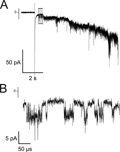
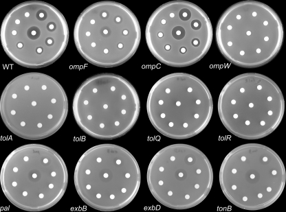
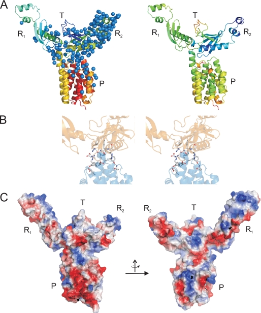
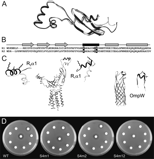
Colicins are plasmid-encoded toxic proteins produced by Escherichia coli strains to kill other E. coli strains that lack the corresponding immunity protein. Colicins intrude into the host cell by exploiting existing transport, diffusion, or efflux systems. We have traced the way colicin S4 takes to execute its function and show that it interacts specifically with OmpW, OmpF, and the Tol system before it inserts its pore-forming domain into the cytoplasmic membrane. The common structural architecture of colicins comprises a translocation, a receptor-binding, and an activity domain. We have solved the crystal structure of colicin S4 to a resolution of 2.5 A, which shows a remarkably compact domain arrangement of four independent domains, including a unique domain duplication of the receptor-binding domain. Finally, we have determined the residues responsible for binding to the receptor OmpW by mutating exposed charged residues in one or both receptor-binding domains.


Figures

FIGURE 1.
FIGURE 1.
A, structural features of the different groups of colicin pores. The pore-forming domains of colicins S4, E1, and Ia shown in the top and side views; the hydrophobic hairpin is drawn in red. The major differences between the colicin groups are the length of the hydrophobic hairpin (see also B), the angle between Pα1 and Pα2 (colicin S4, 5°; colicin E1, 34°; and colicin Ia, 37 °), and the position of the hairpin comprising Pα5 and the loop connection to Pα6. B, superimposition of hydrophobic hairpins. Left, colicin S4 (group PI, in red) versus colicin E1 (group PIIa, in pink); right, colicin E1 (group PIIa, in pink) versus colicin Ia (group PIIb, in purple). C, alignment of the pore-forming domains of colicins. Colicins are grouped according to sequence similarity. Groups PI and PIIa/b differ by several insertions and deletions, whereas groups PIIa and PIIb are highly similar and differ only in several conserved positions, affecting the overall charge and isoelectric point of the domains. Moreover, the structure of colicin Ia contains four very short β-strands where colicin E1 only has unstructured loops.
FIGURE 2.
FIGURE 2.
Single channel conductance measurements. A shows the insertion of numerous channels just after injection of the protein. B shows the action of a single channel flickering at a high frequency, prior to permanent opening. The trace was recorded at -100 mV. The buffer conditions were 1 m KCl and 20 mm MES (pH 6.0).
FIGURE 3.
FIGURE 3.
Colicin S4 sensitivity of selected E. coli strains. The white filter platelets were soaked with colicin S4 (10 μg, middle platelet;1 μg, upper right platelet; continuing clockwise to 10-7 μg). The ΔompW strain shows full resistance. The ΔompF strain shows only partial resistance, indicating that other porins might be used alternatively for the translocation process. The ΔompC strain shows hypersensitivity, probably due to increased ompF expression levels. The ΔtolA, ΔtolB, ΔtolQ, and ΔtolR strains show full resistance, and Δpal shows partial resistance, indicating the Tol-dependent translocation of colicin S4. However, the ΔtonB, ΔexbB, and ΔexbD strains also show partial resistance. WT, wild-type.
FIGURE 4.
FIGURE 4.
Structure of colicin S4. A, stereo view of the complete structure. B, annotation of domains and secondary structure elements: translocation domain (T), receptor-binding domain 1 (R1), receptor-binding domain 2 (R2), and pore-forming domain (P).
FIGURE 5.
FIGURE 5.
Structure of colicin S4. A: left, hydration of colicin S4 in the crystal arrangement. Water molecules are represented by blue balls. Note the significant difference in hydration between domains R1 and R2. Right, structure of colicin S4. B: network of hydrophobic and electrostatic interactions between domain R2 and the pore-forming domain (stereo view). C: electrostatic characterization of colicin S4. Blue, positively charged surface; red, negatively charged surface.
FIGURE 6.
FIGURE 6.
Receptor-binding domain mutants. A, superimposition of the two receptor-binding domains. R1 is shown in black, and R2 is shown in light gray. B, alignment of the two receptor-binding domains. The mutated residues are highlighted in black (positive charge) and gray (negative charge). C, location of the α-helices (black) that were mutated to interfere with OmpW binding. The mutated residues Lys163, Glu170, and Lys171 in R1, and Lys250, Asp257, and Lys258 in R2 are shown in stick representation. D, sensitivity assay. Colicin S4 was spotted on filter platelets in decreasing concentrations (see Fig. 3). The wild-type (WT) E. coli strain is resistant to the colicin mutant S4m12, in which both domains are modified; it is partially resistant to the two colicin mutants S4m1 and S4m2, in which only one domain is changed, and is fully sensitive to control colicin S4.

References

    1. Riley, M. A., and Wertz, J. E. (2002) Annu. Rev. Microbiol. 56 117-137 - PubMed
    1. Cascales, E., Buchanan, S. K., Duche, D., Kleanthous, C., Lloubes, R., Postle, K., Riley, M., Slatin, S., and Cavard, D. (2007) Microbiol. Mol. Biol. Rev. 71 158-229 - PMC - PubMed
    1. Gillor, O., Vriezen, J. A., and Riley, M. A. (2008) Microbiology (Read.) 154 1783-1792 - PMC - PubMed
    1. Lakey, J. H., and Slatin, S. L. (2001) Curr. Top. Microbiol. Immunol. 257 131-161 - PubMed
    1. de Zamaroczy, M., and Buckingham, R. H. (2002) Biochimie (Paris) 84 423-432 - PubMed

Publication types

MeSH terms

Associated data

LinkOut - more resources