. 2009 Apr;19(4):344-55.
doi: 10.1093/glycob/cwn137. Epub 2008 Dec 4.

Atomic-resolution conformational analysis of the GM3 ganglioside in a lipid bilayer and its implications for ganglioside-protein recognition at membrane surfaces

Affiliations

Atomic-resolution conformational analysis of the GM3 ganglioside in a lipid bilayer and its implications for ganglioside-protein recognition at membrane surfaces

Mari L DeMarco et al. Glycobiology. 2009 Apr.

Abstract

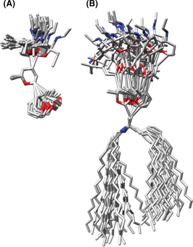
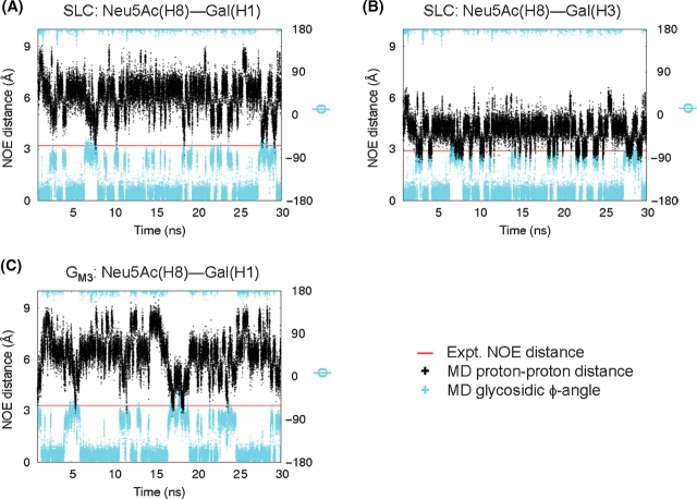
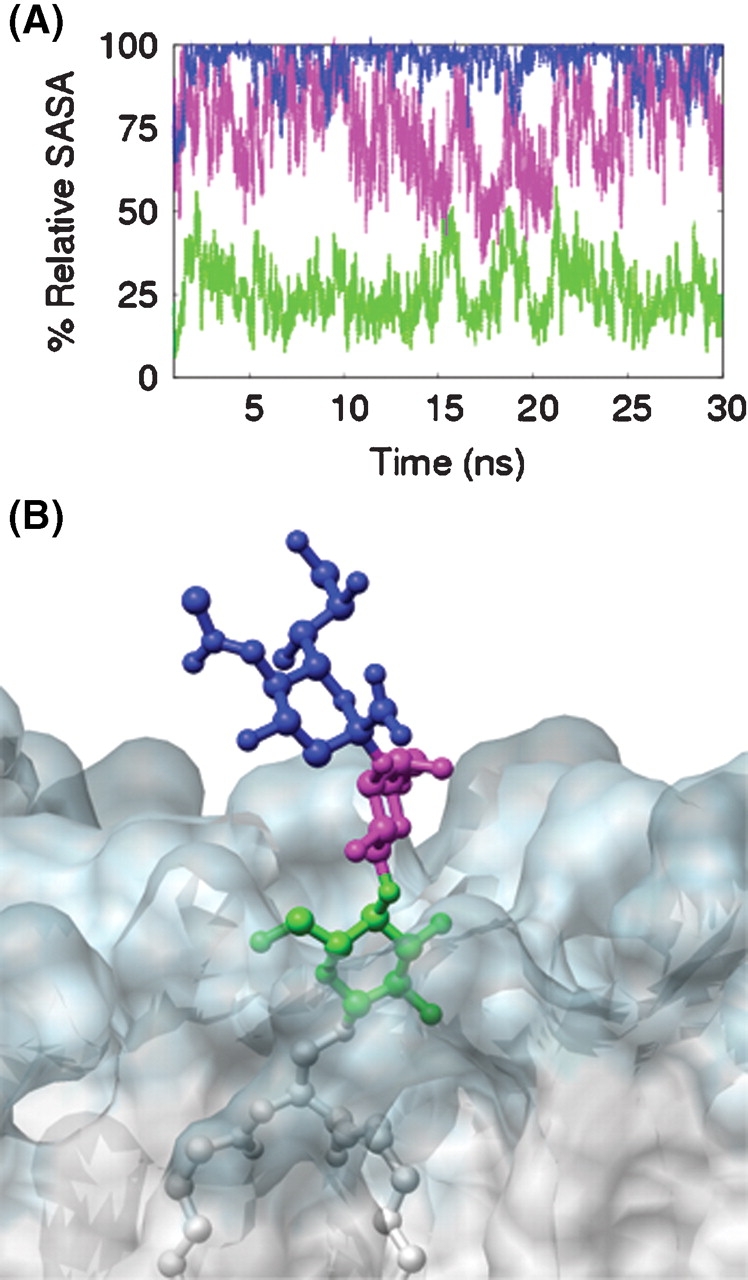
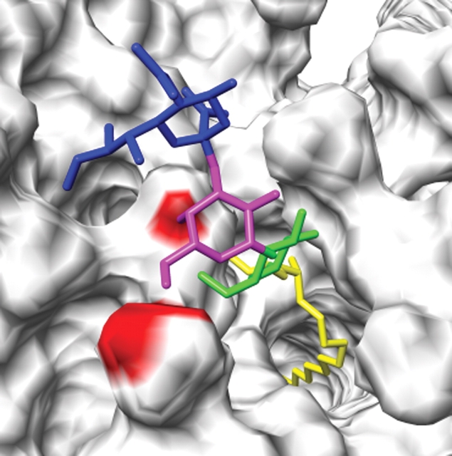
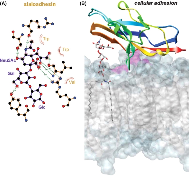
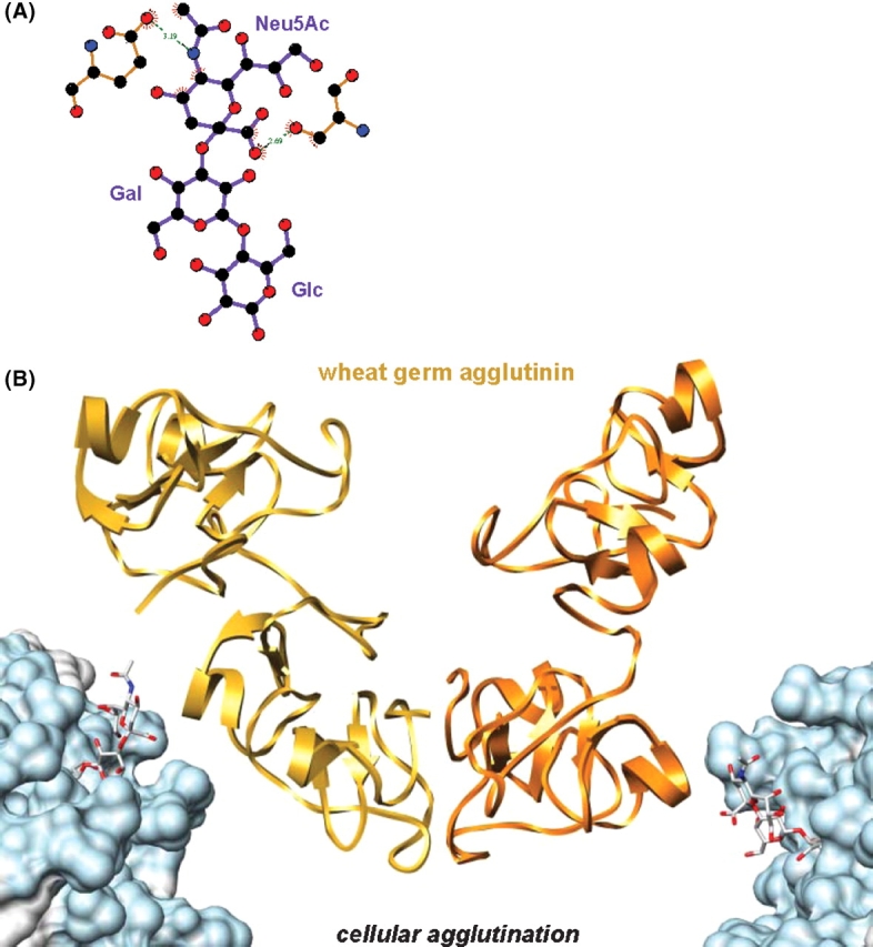
Eukaryotic cells depend on external surface markers, such as gangliosides, to recognize and bind various other molecules as part of normal growth and maturation. The localization of gangliosides in the outer leaflet of the plasma membrane, also make them targets for pathogens trying to invade the host cells. Since ganglioside-mediated interactions are critical to both beneficial and pathological processes, much effort has been directed at determining the 3D structures of their carbohydrate head groups; however, technical difficulties have generally prevented the characterization of the head group in intact membrane-bound gangliosides. Determining the 3D structure and presentation of gangliosides at the surface of membranes is important in understanding how cells interact with their local environment. Here, we employ all-atom explicit solvent molecular dynamics (MD) simulations, using the GLYCAM06 force field, to model the conformation and dynamics of ganglioside G(M3) (alpha-Neu5Ac-(2-3)-beta-Gal-(1-4)-beta-Glc-ceramide) in a DMPC lipid bilayer. By comparison with MD simulations of the carbohydrate head-group fragment of G(M3) alone, it was possible to quantify and characterize the extent of changes in head-group presentation and dynamics associated with membrane anchoring. The accuracy of data from the MD simulations was determined by comparison to NMR and crystallographic data for the head group in solution and for G(M3) in membrane-mimicking environments. The experimentally consistent model of G(M3), in a lipid bilayer, was then used to model the recognition of G(M3) at the cell surface by known protein receptors.


Figures

Fig. 1
Fig. 1
(A) Snapshots from the simulation of SLC superimposed on the ring atoms of the central Gal residue. (B) Snapshots from the simulation of GM3 superimposed on the five nonhydrogen atoms of ceramide closest to Glc. Snapshots are shown at 1 ns intervals, with all hydrogen and oxygen atoms (except for ring oxygens) removed for clarity.
Fig. 2
Fig. 2
Bilayer thickness parameters, calculated from the GM3 simulation, compared to experimental values. The dl values (MD and exp) are shown in the upper two traces and the dhc (MD and exp) in the lower two traces.
Fig. 3
Fig. 3
The population density of glycosidic torsion angle pairs in the simulations of GM3 (A and B) and SLC (C and D). For comparison, glycosidic torsion angles of carbohydrates from the PDB that contain either (E) α-Neu5Ac-(2-3)-β-Gal or (F) β-Gal-(1-4)-β-Glc linkages are shown.
Fig. 4
Fig. 4
Running averages of the scalar 3J-coupling constants (labeled as in Table II) calculated from simulations of (A) GM3 and (B) SLC, which converge to within the experimental values for SLC in solution.
Fig. 5
Fig. 5
Interresidue contacts for which there were significant differences between NOE distances calculated from experiment () (Siebert et al. 1992) and the average interproton distances computed from MD (+). In the simulations of SLC (A and B) and GM3 (C), rotations about the glycosidic bond bring H8 of Neu5Ac into closer proximity to H1 and H3 protons on Gal, as demonstrated by the correlation between the MD NOE distance (+) and the glycosidic ϕ angle (+) for the Neu5Ac–Gal linkage.
Fig. 6
Fig. 6
Changes in the accessibility of the carbohydrate epitope when membrane bound. (A) Relative SASA (%) values for the Neu5Ac (blue), Gal (magenta), and Glc (green) residues of GM3 relative to the ensemble averaged SASA calculated for the same residues from the SLC simulation. (B) 20 ns snapshot taken perpendicular to the plane of the bilayer (transparent space filling mode with hydrophilic region in blue and hydrophobic region in white) near the head group of GM3.
Fig. 7
Fig. 7
Membrane anchoring can restrict conformational space accessible to GM3 relative to SLC. Steric clashes between GM3 (7 ns snapshot) and DMPC molecules when GM3 (Neu5Ac, Gal, Glc, ceramide) is forced into the minor conformer populated by SLC.
Fig. 8
Fig. 8
Cellular adhesion. (A) Sialoadhesin-SLC interactions (1qfo). (B) Crystal structure of sialoadhesin–SLC complex (SLC not shown) rigidly docked to a snapshot of the GM3-DMPC bilayer simulation. The amphipathic Glc-ceramide portion of the ganglioside is found within the DMPC bilayer and is aligned with the bilayer's hydrophilic (light blue) and hydrophobic (gray) regions. Clashes occurring between sialoadhesin and DMPC molecules are shown in magenta.
Fig. 9
Fig. 9
Cell agglutination. (A) Wheat germ agglutinin–SLC interactions (2wgc). (B) The crystal structure of wheat germ agglutinin dimer–SLC complex (SLC not shown) rigidly docked to snapshots of the GM3-DMPC bilayer simulation. Colored as in Figure 8.

References

    1. Airenne TT, Kidron H, Nymalm Y, Nylund M, West G, Mattjus P, Salminen TA. Structural evidence for adaptive ligand binding of glycolipid transfer protein. J Mol Biol. 2006;355:224. - PubMed
    1. Alving CR, Richards RL. Immune reactivities of antibodies against glycolipids: I. Properties of anti-galactocerebroside antibodies purified by a novel technique of affinity binding to liposomes. Immunochemistry. 1977;14:373–381. - PubMed
    1. Ariga T, Yoshida T, Mimori T, Yu RK. Autoantibodies against Forssman glycolipids in Graves’ disease and Hashimoto's thyroiditis. Clin Exp Immunol. 1991;86:483–488. - PMC - PubMed
    1. Aubin Y, Ito Y, Paulson JC, Prestegard JH. Structure and dynamics of the sialic acid moiety of GM3-ganglioside at the surface of a magnetically oriented membrane. Biochemistry. 1993;32:13405–13413. - PubMed
    1. Basma M, Sundara S, Calgan D, Vernali T, Woods RJ. Solvated ensemble averaging in the calculation of partial atomic charges. J Comput Chem. 2001;22:1125–1137. - PMC - PubMed

Publication types

MeSH terms