. 2009 Feb;37(2):506-15.
doi: 10.1093/nar/gkn962. Epub 2008 Dec 4.

An affinity-based scoring scheme for predicting DNA-binding activities of modularly assembled zinc-finger proteins

Affiliations

An affinity-based scoring scheme for predicting DNA-binding activities of modularly assembled zinc-finger proteins

Jeffry D Sander et al. Nucleic Acids Res. 2009 Feb.

Abstract

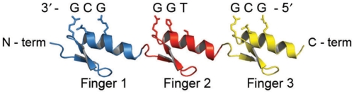
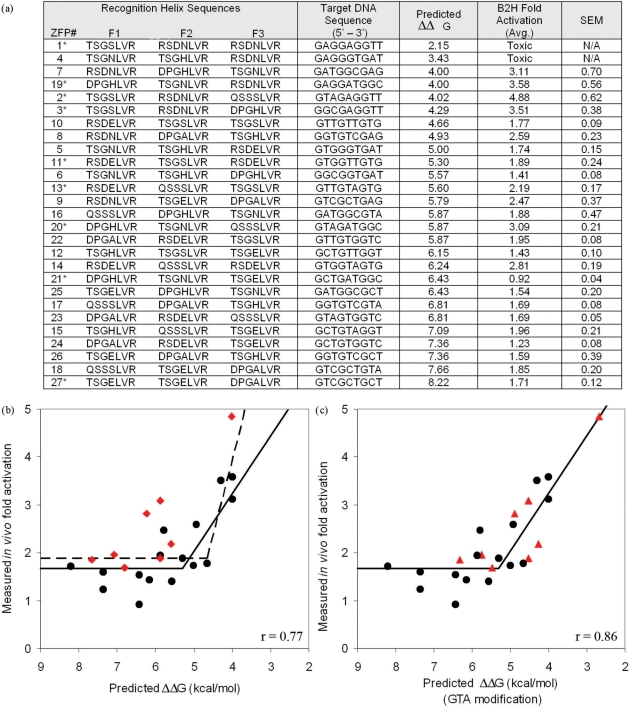
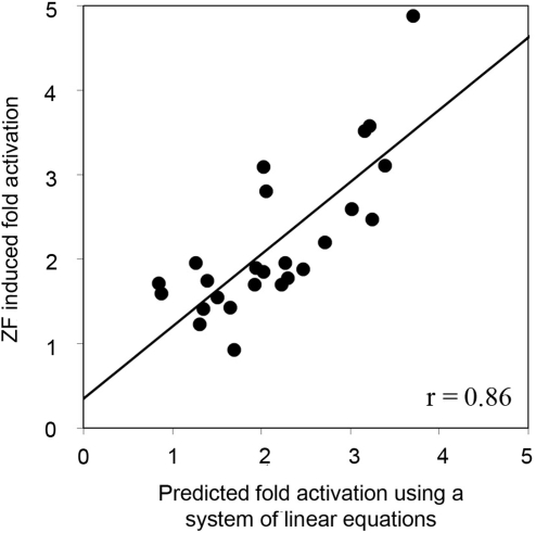
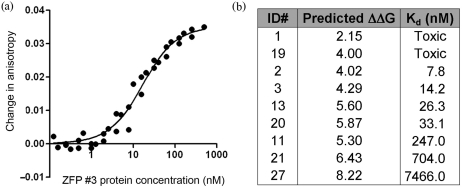
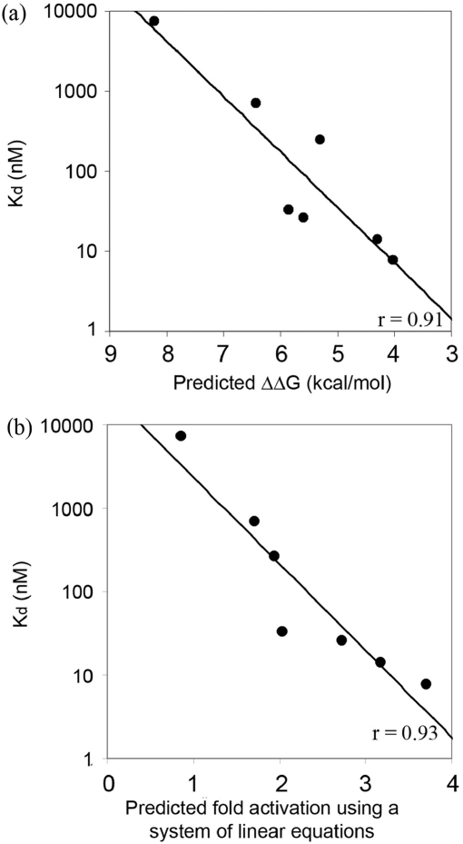
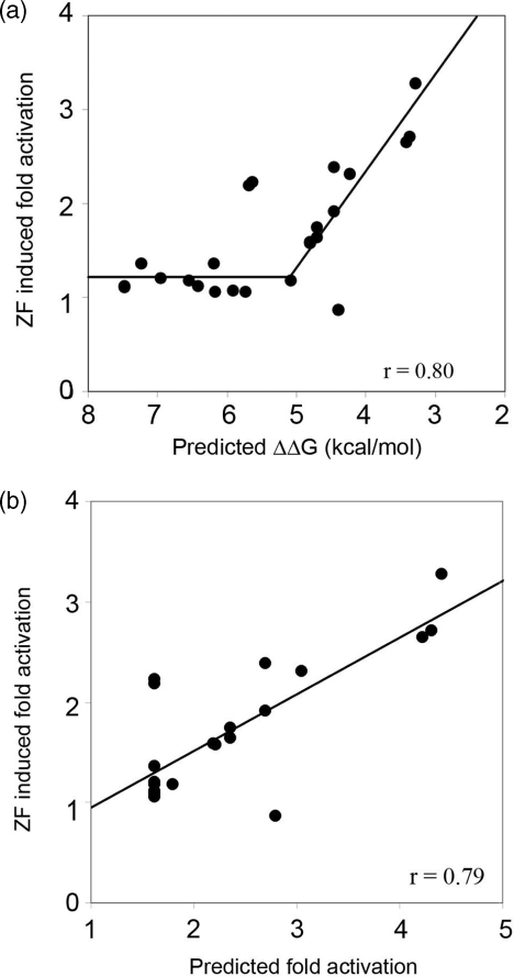
Zinc-finger proteins (ZFPs) have long been recognized for their potential to manipulate genetic information because they can be engineered to bind novel DNA targets. Individual zinc-finger domains (ZFDs) bind specific DNA triplet sequences; their apparent modularity has led some groups to propose methods that allow virtually any desired DNA motif to be targeted in vitro. In practice, however, ZFPs engineered using this 'modular assembly' approach do not always function well in vivo. Here we report a modular assembly scoring strategy that both identifies combinations of modules least likely to function efficiently in vivo and provides accurate estimates of their relative binding affinities in vitro. Predicted binding affinities for 53 'three-finger' ZFPs, computed based on energy contributions of the constituent modules, were highly correlated (r = 0.80) with activity levels measured in bacterial two-hybrid assays. Moreover, K(d) values for seven modularly assembled ZFPs and their intended targets, measured using fluorescence anisotropy, were also highly correlated with predictions (r = 0.91). We propose that success rates for ZFP modular assembly can be significantly improved by exploiting the score-based strategy described here.


Figures

Figure 1.
Figure 1.
A three-finger ZFP with its DNA target site. A ZFP consisting of three adjacent ZFDs binds its target DNA through contacts between the amino acids of the DNA recognition helices and consecutive nucleotides in the DNA. The protein chain is drawn in the N- to C-terminal direction and the DNA target in the 3′–5′ direction. Note that an ‘unnatural’ extended array is shown to better illustrate the critical amino acid/nucleotide contacts. Structure diagrams were generated using PyMol (http://www.pymol.org).
Figure 2.
Figure 2.
Predicted binding energies are highly correlated with in vivo activity in a B2H assay. Twenty-seven three-module ZFPs were designed by modular assembly to span a broad range of predicted binding affinities. (a) DNA recognition helix sequences, DNA sequence targets, predicted energies, measured fold-activation in the B2H assay, and standard error of the mean are listed for each construct. Entries are sorted from lowest to highest predicted ΔΔG. Constructs marked with an asterisk were also tested in vitro (Figure 4). (b) ZFP activities in the B2H assay are plotted versus predicted energies. The two constructs with highest predicted affinities were toxic to their host cells and therefore could not be included. Points shown as red diamonds correspond to proteins containing the GTA-specific QSSSLVR module (see text). Best-fit lines from a segmental linear regression model using all points (dashed line, r = 0.77), or excluding red points (solid line, r = 0.86) are shown. (c) Same as (b), except that values indicated by red triangles were adjusted assuming a binding affinity of 2.5 nM, rather than 25 nM, for the GTA-specific QSSSLVR module; this increases the correlation coefficient to 0.86 (see text for details).
Figure 3.
Figure 3.
ZFDs contribute additively to B2H activity, independent of context and position. For each ZF protein, the expected contribution to B2H activity from each of its component modules was estimated by solving a system of linear equations representing the other 24 proteins (see text). Comparison of actual versus predicted B2H activity (expressed as relative fold-activation in the B2H assay) reveals a high correlation (r = 0.86).
Figure 4.
Figure 4.
Determining binding affinity constants using fluorescence anisotropy. (a) A representative in vitro binding isotherm obtained using FA. Data points for each ZFP were collected using three separate purified protein preparations, each assayed for binding activity on a different day. Curve fitting was performed using Prism. (b) Kd values for seven modularly assembled ZFPs, determined in FA experiments. Note that two ZFPs were toxic to host cells, preventing purification of proteins in quantities required for in vitro analysis.
Figure 5.
Figure 5.
In vitro affinity constants for ZFPs are highly correlated with predictions. Using affinity constants for seven ZFPs determined by FA (Figure 4b), log(Kd) is plotted against: (a) predicted energy, expressed as ΔΔG in kcal/mol (r = 0.91) and (b) predicted B2H activity based on a leave-one-out system of linear equations analysis (r = 0.93).
Figure 6.
Figure 6.
Predicted ZFP performance agrees with in vivo activity for an independently generated set of ZFPs. Data shown are for 24 of 26 ZFPs containing characterized GNN or TGG modules, constructed and evaluated by Ramirez et al. (25) (a) A segmental linear regression model provides an excellent fit of in vivo ZF-induced fold activation measured in the B2H assay with predicted binding energies (r = 0.80). (b) ZF-induced fold activation values measured in the B2H assay for 24 ZFPs from Ramirez et al. (25) are also highly correlated (r = 0.79) with predicted fold-activation levels calculated based on a scoring function derived from the segmental linear regression model fit for the 25 ZFPs shown in Figure 2a (see text for details). Note: predictions for 2 of 26 ZFPs containing characterized GNN or TGG modules from Ramirez et al. (25) were considered outliers (values were outside the range included in these graphs); they were not included in the regression analysis.

Comment in

References

    1. Durai S, Mani M, Kandavelou K, Wu J, Porteus MH, Chandrasegaran S. Zinc finger nucleases: custom-designed molecular scissors for genome engineering of plant and mammalian cells. Nucleic Acids Res. 2005;33:5978–5990. - PMC - PubMed
    1. Klug A. Towards therapeutic applications of engineered zinc finger proteins. FEBS Lett. 2005;579:892–894. - PubMed
    1. Porteus MH, Carroll D. Gene targeting using zinc finger nucleases. Nat. Biotechnol. 2005;23:967–973. - PubMed
    1. Wu J, Kandavelou K, Chandrasegaran S. Custom-designed zinc finger nucleases: what is next? Cell. Mol. Life Sci. 2007;64:2933–2944. - PMC - PubMed
    1. Cathomen T, Joung JK. Zinc-finger nucleases: the next generation emerges. Mol. Ther. 2008;16:1200–1207. - PubMed

Publication types

Substances