. 2009 Feb;37(2):493-505.
doi: 10.1093/nar/gkn961. Epub 2008 Dec 4.

UHRF1 binds G9a and participates in p21 transcriptional regulation in mammalian cells

Affiliations

UHRF1 binds G9a and participates in p21 transcriptional regulation in mammalian cells

Jong Kyong Kim et al. Nucleic Acids Res. 2009 Feb.

Abstract

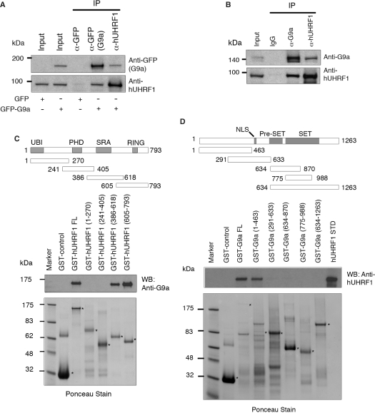
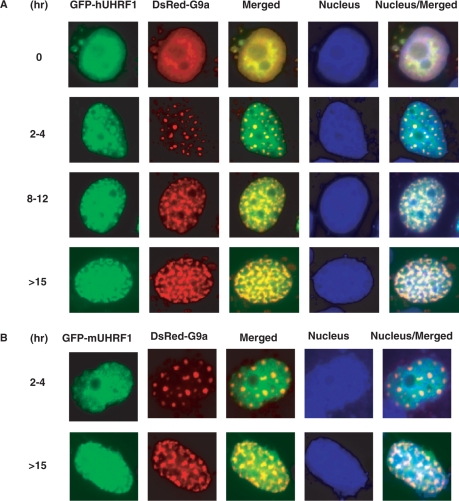
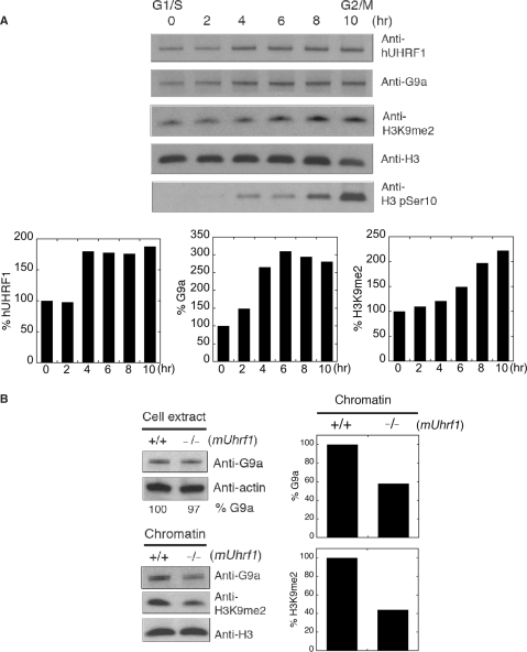
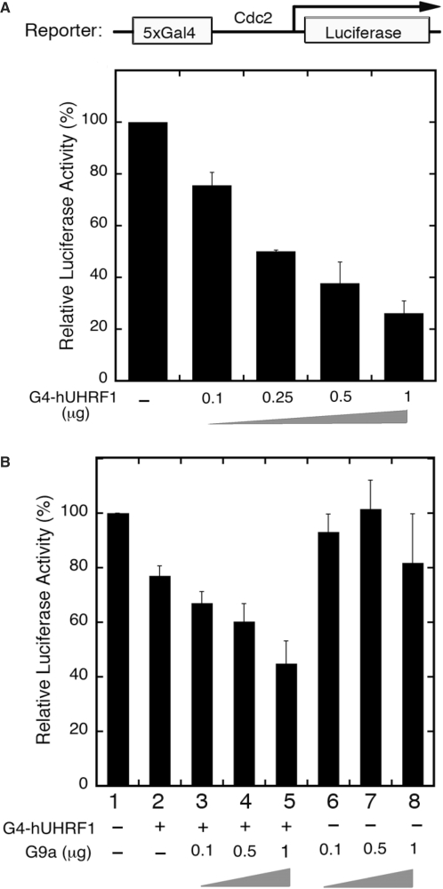
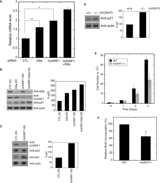
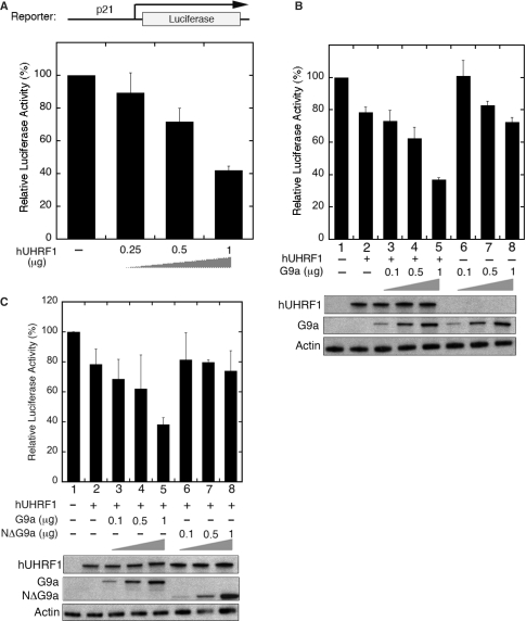
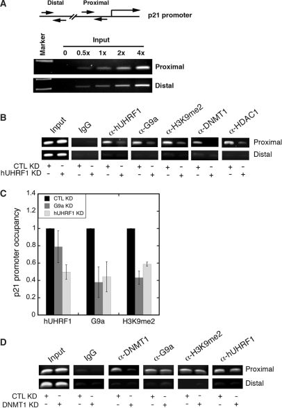
UHRF1 (ubiquitin-like, containing PHD and RING finger domains 1) is a multi-domain protein associated with cellular proliferation and epigenetic regulation. The UHRF1 binds to methylated CpG dinucleotides and recruits transcriptional repressors DNA methyltransferase 1 (DNMT1) and histone deacetylase 1 (HDAC1) through its distinct domains. However, the molecular basis of UHRF1-mediated transcriptional regulation via chromatin modifications is yet to be fully understood. Here we show that UHRF1 binds histone lysine methyltransferase G9a, and both are co-localized in the nucleus in a cell-cycle-dependent manner. Concurrent with the cell-cycle progression, gradual deposition of UHRF1 and G9a was observed, which mirrored H3K9me2 accumulation on chromatin. Murine Uhrf1-null embryonic stem (ES) cells displayed a reduced amount of G9a and H3K9me2 on chromatin. UHRF1 recruited and cooperated with G9a to inhibit the p21 promoter activity, which correlated with the elevated p21 protein level in both human UHRF1 siRNA-transfected HeLa cells and murine Uhrf1-null ES cells. Furthermore, endogenous p21 promoter remained bound to UHRF1, G9a, DNMT1 and HDAC1, and knockdown of UHRF1 impaired the association of all three chromatin modifiers with the promoter. Thus, our results suggest that UHRF1 may serve as a focal point of transcriptional regulation mediated by G9a and other chromatin modification enzymes.


Figures

Figure 1.
Figure 1.
Physical interaction of hUHRF1 and G9a. (A) Coimmunoprecipitation of hUHRF1 and G9a in cell extracts from COS-7 cells transfected with GFP control or GFP-G9a. The antibodies used for immunoprecipitation (IP) are indicated at the top of the panel. Western-blot analysis of immunoprecipitates was performed with antibodies as indicated. The input shows 2% of each lysate. (B) Coimmunoprecipitation of endogenous hUHRF1 and G9a in HEK293 cell extracts. HEK293 cells were synchronized to late S phase by serum starvation for 20 h and the subsequent release into 10% FBS-containing medium for 15 h before cell harvest. Antibodies used for immunoprecipitation and western blot are shown. The input represents 2% of each lysate. (C) Direct binding of hUHRF1 to G9a and mapping of the G9a-binding region on hUHRF1 using GST fusions of hUHRF1 fragments. Various domains of hUHRF1 are indicated along with a schematic presentation of the GST fusion constructs marked with amino acid numbers. The blot from GST pull-down assay was probed with anti-G9a antibody and stained with Ponceau solution to visualize the transferred proteins. Positions of fusion proteins are marked with asterisks. (D) Mapping of hUHRF1-binding region on G9a. Schematic diagram of the various GST-fusion constructs of G9a is shown with amino acid numbers. Western blot with anti-hUHRF1 antibody is shown along with the corresponding Ponceau-stained membrane.
Figure 2.
Figure 2.
Colocalization of UHRF1 and G9a. (A) Subnuclear localization of GFP-hUHRF1 and DsRed-G9a transiently expressed in COS-7 cells. Cells were synchronized with aphidicolin and released from G1 arrest for a given number of hours through S phase. Nuclei were visualized with Hoechst stain. (B) Colocalization of GFP-mUHRF1 and DsRed-G9a in COS-7 cells at given hours of release from synchronization.
Figure 3.
Figure 3.
UHRF1 affects chromatin association of G9a. (A) Concurrent loading of hUHRF1 and G9a onto chromatin during S phase. HeLa cells were synchronized by double aphidicolin/thymidine block and released to regular medium for hours indicated at the top of the panel. The chromatin fractions at each time point were used for western-blot analysis with antibodies indicated. Anti-H3 and H3 phopho-Ser10 (H3 pSer10) antibodies were used for a loading control and mitosis marker, respectively. Densitometric scans of hUHRF1, G9a and H3K9me2 levels in each chromatin fraction are shown after normalization by the corresponding H3 level. (B) Impaired chromatin association of G9a in the absence of mUHRF1. Cell extracts and chromatin were prepared from wild type (+/+) and mUhrf1-null (−/−) ES (ES) cells and used to detect the presence of G9a and dimethylated H3K9 (H3K9me2) with antibodies indicated. The relative G9a level in cell extracts is shown as percentage G9a at the bottom of the panel by a densitometric analysis after normalization by the actin-loading control. Normalized densitometric scans of G9a and H3K9me2 levels in chromatin are shown in graphs.
Figure 4.
Figure 4.
hUHRF1 cooperates with G9a to enhance transcriptional repression. (A) Transcriptional repression of luciferase reporter gene by hUHRF1. COS-7 cells were cotransfected with a Gal4-driven luciferase reporter (2 μg) and increasing amounts of Gal4DBD-hUHRF1 (G4-hUHRF1). Luciferase activity was measured 48 h post-transfection and represented by the means ± SD of duplicate determinations from three independent experiments. (B) Enhanced transcriptional repression by hUHRF1-mediated G9a recruitment. Luciferase activities were measured after 48 h of transfection using COS-7 cells cotransfected with the same reporter in (A), increasing amounts of EGFP-G9a (0.1–1 μg) and with or without a constant amount (0.1 μg) of G4-hUHRF1. The data represent the means ± SD of duplicate determinations from four separate experiments.
Figure 5.
Figure 5.
hUHRF1 suppresses p21 expression in cooperation with G9a. (A) Quantitative RT–PCR analysis of p21 expression. Total RNA was isolated from HeLa cells transfected with siRNAs as indicated. The Q-PCR data normalized by GAPDH control are shown as the means ± SD of triplicate determinations from four independent experiments. Statistical significance of the differences among the groups was determined by Student's t-test. *P < 0.05; **P < 0.01. (B) Western blot analysis of p21 expression in siRNA-mediated KD HeLa cells. After each KD, as indicated at the top of the panel, cell extracts were used for detection of p21. The densitometric scan of p21 expression is shown at the right. (C) Western blot analysis of p53 and p21 expression in COS-7 cells after siRNA-mediated KD of hUHRF1. The densitometric scan of p21 expression is shown at the right. (D) Enhanced p21 expression in mUhrf1−/− ES cells. The blot for p21 is shown along with the densitometric analysis. (E) Growth of wild type (+/+) and mUhrf1-null (−/−) ES cells. Cell growth was monitored over 3 days after plating, and the data represent the means ± SD of six replicates. (F) BrdU incorporation of wild-type (+/+) and mUhrf1-null (−/−) ES cells. After 24 h incubation, BrdU labeling was performed for 2 h and determined by a colorimetric assay. The data represent the means ± SD of three separate experiments. Statistical significance of the difference between the groups was determined by Student's t-test. *P < 0.05.
Figure 6.
Figure 6.
hUHRF1 cooperates with G9a to enhance the transcriptional repression of p21 promoter. (A) hUHRF1-mediated transcriptional repression of the p21 promoter-luciferase reporter. COS-7 cells were cotransfected with the reporter (2 μg) and increasing amounts of EGFP-hUHRF1 as indicated at the bottom of the panel. (B) Enhanced transcriptional repression by G9a in the presence of exogenous hUHRF1. Luciferase activities were measured from COS-7 cells cotransfected with the same reporter described earlier and increasing amounts of EGFP-G9a (0.1–1 μg) with or without a constant amount (0.4 μg) of EGFP-hUHRF1. The luciferase assays were performed as described in Figure 4, and the data represent the means ± SD of duplicate determinations from three separate experiments. Western blot analyses of hUHRF1 and G9a expression by anti-GFP antibody are shown for each cotransfection group. (C) Loss of interaction between UHRF1 and G9a abolishes the UHRF1/G9a-mediated repression of p21 promoter. Reporter assays were performed as described in (B), using the wild-type G9a plasmid (EGFP-G9a) and its deletion mutant lacking the N-terminal UHRF1-interacting region (EGFP-NΔG9a). Expression of hUHRF1 and G9a/NΔG9a is shown by western blot analyses with anti-GFP antibody.
Figure 7.
Figure 7.
hUHRF1 recruits G9a and other chromatin modification enzymes to p21 promoter. (A) Linearity of PCR amplification using primer sets for proximal (−385 to −240) and distal (−4164 to −3959) regions of p21 promoter with increasing amount of input DNA. (B) ChIP analysis of p21 promoter after KD of hUHRF1. HeLa cells were transfected with either control siRNA (CTL KD) or hUHRF1 siRNA (hUHRF1 KD). Using the chromatin isolated from the KD cells, ChIP was performed to detect the proteins or histone modification as indicated at the top of the panel. 5% input is shown. (C) Quantitative ChIP analysis for relative p21 promoter occupancy of hUHRF1, G9a and dimethylated H3K9 (H3K9me2) after KD of hUHRF1 or G9a. Q-PCR data of each group were normalized to its input as % input. The relative p21 promoter occupancy of hUHRF1 or G9a KD samples represents the fold change in percentage input over that of the CTL KD. Error bars indicate standard deviation of three independent experiments. (D) ChIP analysis of p21 promoter after KD of DNMT1. HeLa cells were transfected with either control siRNA (CTL KD) or DNMT1 siRNA (DNMT1 KD). CHIP was performed as described in (B) and 5% input is shown.

References

    1. Bronner C, Achour M, Arima Y, Chataigneau T, Saya H, Schini-Kerth VB. The UHRF family: oncogenes that are drugable targets for cancer therapy in the near future? Pharmacol. Ther. 2007;115:419–434. - PubMed
    1. Hopfner R, Mousli M, Jeltsch JM, Voulgaris A, Lutz Y, Marin C, Bellocq JP, Oudet P, Bronner C. ICBP90, a novel human CCAAT binding protein, involved in the regulation of topoisomerase IIalpha expression. Cancer Res. 2000;60:121–128. - PubMed
    1. Muto M, Utsuyama M, Horiguchi T, Kubo E, Sado T, Hirokawa K. The characterization of the monoclonal antibody Th-10a, specific for a nuclear protein appearing in the S phase of the cell cycle in normal thymocytes and its unregulated expression in lymphoma cell lines. Cell Prolif. 1995;28:645–657. - PubMed
    1. Karagianni P, Amazit L, Qin J, Wong J. ICBP90, a novel methyl K9 H3 binding protein linking protein ubiquitination with heterochromatin formation. Mol. Cell Biol. 2008;28:705–717. - PMC - PubMed
    1. Citterio E, Papait R, Nicassio F, Vecchi M, Gomiero P, Mantovani R, Di Fiore PP, Bonapace IM. Np95 is a histone-binding protein endowed with ubiquitin ligase activity. Mol. Cell Biol. 2004;24:2526–2535. - PMC - PubMed

Publication types

MeSH terms