. 2008 Dec 1;22(23):3292-307.
doi: 10.1101/gad.1734608.

UNC-51/ATG1 kinase regulates axonal transport by mediating motor-cargo assembly

Affiliations

UNC-51/ATG1 kinase regulates axonal transport by mediating motor-cargo assembly

Hirofumi Toda et al. Genes Dev. .

Abstract

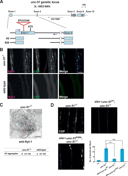
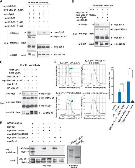
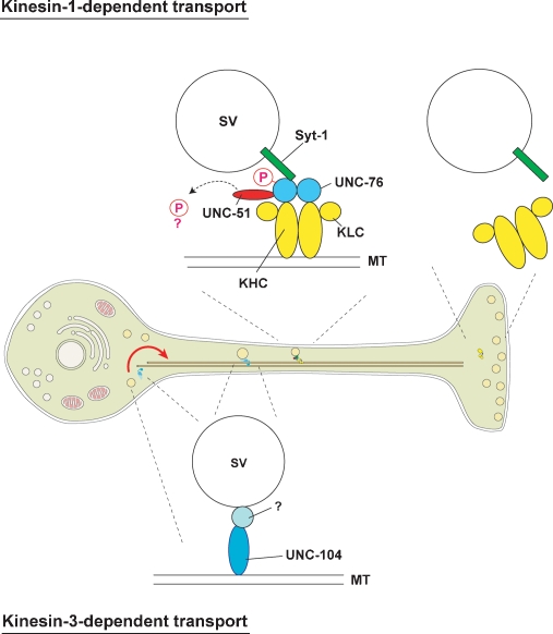
Axonal transport mediated by microtubule-dependent motors is vital for neuronal function and viability. Selective sets of cargoes, including macromolecules and organelles, are transported long range along axons to specific destinations. Despite intensive studies focusing on the motor machinery, the regulatory mechanisms that control motor-cargo assembly are not well understood. Here we show that UNC-51/ATG1 kinase regulates the interaction between synaptic vesicles and motor complexes during transport in Drosophila. UNC-51 binds UNC-76, a kinesin heavy chain (KHC) adaptor protein. Loss of unc-51 or unc-76 leads to severe axonal transport defects in which synaptic vesicles are segregated from the motor complexes and accumulate along axons. Genetic studies show that unc-51 and unc-76 functionally interact in vivo to regulate axonal transport. UNC-51 phosphorylates UNC-76 on Ser(143), and the phosphorylated UNC-76 binds Synaptotagmin-1, a synaptic vesicle protein, suggesting that motor-cargo interactions are regulated in a phosphorylation-dependent manner. In addition, defective axonal transport in unc-76 mutants is rescued by a phospho-mimetic UNC-76, but not a phospho-defective UNC-76, demonstrating the essential role of UNC-76 Ser(143) phosphorylation in axonal transport. Thus, our data provide insight into axonal transport regulation that depends on the phosphorylation of adaptor proteins.


Figures

Figure 1.
Figure 1.
unc-51 functions in axonal transport. (A) Molecular structures of the unc-51-null mutant alleles. P-element EP(3)3348 is inserted within the first exon of the unc-51 gene, 539 bp upstream of the initiation ATG codon. Two unc-51-null alleles, unc-513 and unc-5125, were generated by imprecise excision. PCR amplification of these unc-51 alleles (blue arrows indicate locations of primers) and subsequent sequencing analyses confirmed that unc-513 has an 804 bp deletion and unc-5125 has a 1787-bp deletion. Both alleles lack the start codon, and did not affect the expression unit of CG17667 located within intron 2. (B) SGNs of unc-51-null mutant (unc-513/unc-5125) or wild-type third instar larvae immunostained with anti-Syt-1 (red) and anti-CSP (green). Syt-1-positive aggregates colocalized with those positive for CSP in unc-51 mutants. Bar, 10 μm. (C) Ultrastructure of SV aggregates in unc-51 mutants. Electron micrograph of a cross-section of an unc-51 mutant SGN immunostained with anti-Syt-1. Arrowheads indicate SVs positive for anti-Syt-1. (Table) The number of SV aggregates (defined as a cluster of >20 SVs per aggregate) was scored in electron-micrographs from unc-51 mutants (n = 34) and wild type (n = 23). Bar, 200 nm. (D) Genetic rescue of axonal transport defect in unc-51 mutants by unc-51 transgene. CSP-positive SV aggregation phenotype in unc-51 mutants was rescued by pan-neuronal expression of unc-51/wild-type transgene (elavc155 > UAS-unc-51WT; unc-513/unc-5125), but not by a kinase-deficient unc-51 (elavc155 > UAS-unc-51K38A; unc-513/unc-5125). Numbers of CSP-positive puncta per 50 μm SGN (mean ± SEM) were plotted for each genotype (graph). (**) Statistically significant rescue (P < 0.01, Student t test). (n.s.) Not significant (P = 0.843). Bar, 10 μm.
Figure 2.
Figure 2.
unc-51 genetically interacts with unc-76 in axonal transport. SGNs of wild type or the indicated mutants immunostained with anti-CSP at the third instar larval stage. Arrows show CSP-positive aggregates. Bar, 20 μm. The number of aggregates found per 50 μm of nerve shaft was scored for each genotype (graph). Statistical significance evaluated by Student t test. (**) P < 0.01. Error bars show ±SEM. Genotypes are w- (A), unc-513/+ (B), unc-76Df(1)107/+ (C), and unc-76Df(1)107/+;unc-513/+ (D).
Figure 3.
Figure 3.
Physical interaction of UNC-51, UNC-76, and KHC. (A) Myc-tagged UNC51 (full-length) and HA-tagged UNC-76 (truncation mutants) were coexpressed in HEK293T cells as indicated. Cell lysates were immunoprecipitated by anti-HA followed by immunoblot with anti-myc. (*) Nonspecific band (22 kDa) detected by anti-HA antibody. (B) UNC-51 binds UNC-76 in vitro. GST alone or GST fused with full-length UNC-76 was produced in E. coli, purified on Glutathione-Sepharose beads, and incubated with myc-tagged UNC-51 C′-terminal domain (amino acids 557–855) produced by in vitro translation (IVT). Proteins that bound to the beads were eluted and analyzed by immunoblot using anti-myc. (C) Schematic representation of the domains involved in UNC-51−UNC-76 binding. Black bars show deletion constructs that bind the other in coimmunoprecipitation assays. Constructs shown in gray did not bind. Binding of all deletion constructs except for UNC-76 (118−340 amino acids) were tested by coimmunoprecipitation assays in A and Supplemental Figure S5, whereas no binding of UNC-76 (118−340 amino acids) with UNC-51 was confirmed by GST pull-down assay (data not shown). The UNC-76 and UNC-51 functional domains are indicated within each protein. (D) UNC-51 forms a complex with KHC via UNC-76. HEK293T cell extracts containing heterologously expressed UNC-51 (full-length), UNC-76 (full-length), and KHC (full-length), as indicated, were analyzed by immunoprecipitation with anti-HA followed by immunoblot with the indicated antibodies.
Figure 4.
Figure 4.
Disorganized localization of SV and motor complexes in unc-51 mutants. (A) SGNs of unc-51 mutants (unc-513/unc-5125) or wild-type third instar larvae immunostained with anti-UNC-76 (red) and anti-Syt-1 (green). Red arrows and arrowheads point to aberrant localization of the indicated proteins. The right panel shows enlarged image (arrowheads, UNC-76; arrows, Syt-1). The numbers of puncta positive for UNC-76 and/or Syt-1 in unc-51 mutant SGNs were scored and the percentages of single-positive or double-positive puncta are shown in the graph. SGNs are oriented from the proximal side (top) toward the distal (bottom). (B) SGNs of unc-51 mutants or wild-type larvae immunostained with anti-UNC-76 (red) and anti-KHC (green). Red arrows point to aberrant localization of the indicated proteins. The right panel shows enlarged image (asterisks, KHC/UNC-76 double-positive aggregates). The numbers of puncta positive for UNC-76 and/or KHC in unc-51 mutant SGNs were scored and the percentages of single-positive or double-positive puncta are shown (graph). (C) SGNs of unc-51 mutants or wild-type larvae immunostained with anti-CSP (red) and anti-KHC (green). Red arrows and arrowheads point to aberrant localization of the indicated proteins. Right panel shows enlarged image (arrowheads, CSP; arrows, KHC). (Graph) The numbers of puncta positive for CSP and/or KHC in unc-51 mutant SGNs were scored and the percentages of single-positive or double-positive puncta are shown. (D) SGNs of unc-51 mutant or wild-type larvae immunostained with anti-CSP (red) and anti-UNC-76 (green) and observed with confocal microscopy (with 100× objective; single optical section taken at 0.7-μm interval). SGNs are oriented from the proximal side (left) toward the distal (right). Areas positive for CSP, UNC-76 or both signals were calculated based on the confocal data using an image analysis program, and schematically represented on the graph. Bars, 10 μm.
Figure 5.
Figure 5.
Phosphorylation of UNC-76 Ser143 by UNC-51 is necessary for axonal transport. (A) In vitro phosphorylation of UNC-76 by UNC-51 kinase. HA-tagged UNC-51 or UNC-51/K38A expressed in HEK293T cells was immunoprecipitated and incubated with His-UNC-76 in the presence of Mg2+ and [γ-32P]-ATP. Phosphoproteins were analyzed by autoradiograph. Black arrow indicates UNC-51 autophosphorylation. Red arrowhead indicates UNC-76 phosphorylation. (B) Myc-tagged UNC-76 and HA-tagged UNC-51 were coexpressed in HEK293T cells. Cell lysates were incubated for an indicated period of time with (+) or without (−) alkaline phosphatase (CIAP) and analyzed by immunoblot using anti-myc. Red bracket indicates mobility shifts of UNC-76. Black bracket indicates nonshifted UNC-76. (C) Myc-tagged wild type and Ser/Thr → Ala mutant UNC-76 were expressed in HEK293T cells with UNC-51 or UNC-51/K38A. Cell lysates were analyzed by immunoblot using anti-myc. Red bracket indicates mobility shifts of UNC-76. (D) Extracts from wild-type and unc-51 mutant larvae were analyzed by immunoblot using an anti-phospho-UNC-76 antibody. Anti-actin and anti-UNC-76 were used as loading controls. (E) Genetic rescue of axonal transport defect in unc-76 mutants by unc-76 transgene. CSP-positive aggregation (arrows) and KHC aggregation (arrowheads) in unc-76 mutants were rescued by unc-76WT (top) or unc-76S143D (middle), but not by unc-76S143D transgene (bottom). Transgene expression was driven by elav. unc-76DF(1)107 hemizygous males were used as unc-76-null mutants. Third instar larvae for each genotype were stained with anti-KHC (green), anti-UNC-76 (red), and anti-CSP (blue). Note the equivalent expression levels of UNC-76 transgenes in VNC and SGN. (**) Statistical significance (P < 0.01, Student t test). (n.s.) Not significant (P = 0.18). Bars: red, 50 μm; white, 20 μm.
Figure 6.
Figure 6.
UNC-51 regulates the affinity between UNC-76 and Syt-1. (A) UNC-51-mediated association of Syt-1 and UNC-76. Myc- or HA-tagged UNC-51 (full-length), UNC-76 (full-length), and Syt-1 (full-length) were coexpressed in HEK293T cells as indicated. Cell lysates were immunoprecipitated by anti-HA followed by immunoblot with anti-myc. (B) Phosphorylation-dependent association of Syt-1 and UNC-76. HA-UNC-76, myc-Syt-1, and myc-UNC-51 or UNC-51/K38A were expressed in HEK293T cells as indicated. Cell lysates were immunoprecipitated with anti-HA. The immunecomplexes were incubated in the presence (+) or absence (−) of alkaline phosphatase (CIAP) and analyzed by immunoblot using anti-myc. (C) Syt-1–UNC-76 interaction does not require Ca2+. HA-UNC-76, myc-Syt-1, and myc-UNC-51 or UNC-51/K38A were expressed in HEK293T cells as indicated. Cell lysates were immunoprecipitated with anti-HA. The immune complexes were incubated in the presence of EGTA or CaCl2 and analyzed by immunoblot using anti-myc. (D) Syt-1–UNC-76 interaction detected by FRET. COS7 cells were transfected with Syt-1-CFP, YFP-UNC-76, YFP-UNC-76/S143D, YFP-UNC-76/S143A, or UNC-51/K38A in combination as indicated. For each combination, initial (red) and fluorescence after acceptor photobleaching spectra (green) were plotted for a range of wavelengths indicated. Increase in donor fluorescence after acceptor bleaching indicates FRET (green arrows). Quantification of FRET (graph). (**) Statistical significance (P < 0.01). (E) KHC/UNC-76/Syt-1 complex formation. GST alone (G) and GST fused with KHC (amino acids 675–975) (GK) were produced in E. coli, purified on Glutathione-Sepharose beads, and incubated with myc-UNC-76 and myc-Syt-1 expressed in HEK293T cells. Proteins that bound to the beads were eluted and analyzed by immunoblot using anti-myc.
Figure 7.
Figure 7.
Schematic model for UNC-51 kinase-mediated motor–cargo assembly Our results suggest a model in which UNC-51 kinase functions in axonal transport via the kinesin-1-dependent pathway. When phosphorylated by UNC-51 kinase, UNC-76, a KHC adaptor and a potential activator of kinesin-1, displays an increased affinity to SV membrane proteins such as Syt-1. UNC-51 likely phosphorylates additional substrates that are essential for axonal transport (dashed arrow). Loss or attenuation of UNC-51 kinase activity would result in a lower affinity of UNC-76 to SV membrane proteins and dissociation of SV cargoes from the motor complexes, as might be the case at NMJ. Kinesin-3 (imac/UNC-104) is also responsible for SV transport in a large fraction and is important for carrying SVs from neuronal soma to axons and synapses (red arrow).

References

    1. Ahantarig A., Chadwell L.V., Terrazas I.B., Garcia C.T., Nazarian J.J., Lee H.K., Lundell M.J., Cassill J.A. Molecular characterization of Pegarn: A Drosophilahomolog of UNC-51 kinase. Mol. Biol. Rep. 2008 doi: 10.1007/s11033-008-9314-4. - DOI - PMC - PubMed
    1. Barkus R.V., Klyachko O., Horiuchi D., Dickson B.J., Saxton W.M. Identification of an axonal Kinesin-3 motor for fast anterograde vesicle transport that facilitates retrograde transport of neuropeptides. Mol. Biol. Cell. 2008;19:274–283. - PMC - PubMed
    1. Blasius T.L., Cai D., Jih G.T., Toret C.P., Verhey K.J. Two binding partners cooperate to activate the molecular motor Kinesin-1. J. Cell Biol. 2007;176:11–17. - PMC - PubMed
    1. Bloom L., Horvitz H.R. The Caenorhabditis elegans gene unc-76 and its human homologs define a new gene family involved in axonal outgrowth and fasciculation. Proc. Natl. Acad. Sci. 1997;94:3414–3419. - PMC - PubMed
    1. Bloom G.S., Richards B.W., Leopold P.L., Ritchey D.M., Brady S.T. GTP γ S inhibits organelle transport along axonal microtubules. J. Cell Biol. 1993;120:467–476. - PMC - PubMed

Publication types

MeSH terms

LinkOut - more resources