. 2008 Dec 5;322(5907):1562-5.
doi: 10.1126/science.1164511.

Maternal alloantigens promote the development of tolerogenic fetal regulatory T cells in utero

Affiliations

Maternal alloantigens promote the development of tolerogenic fetal regulatory T cells in utero

Jeff E Mold et al. Science. .

Abstract

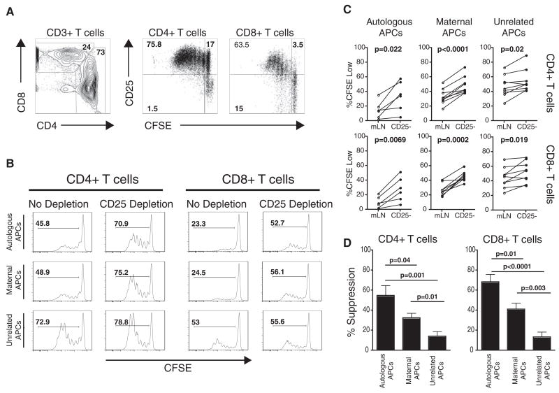
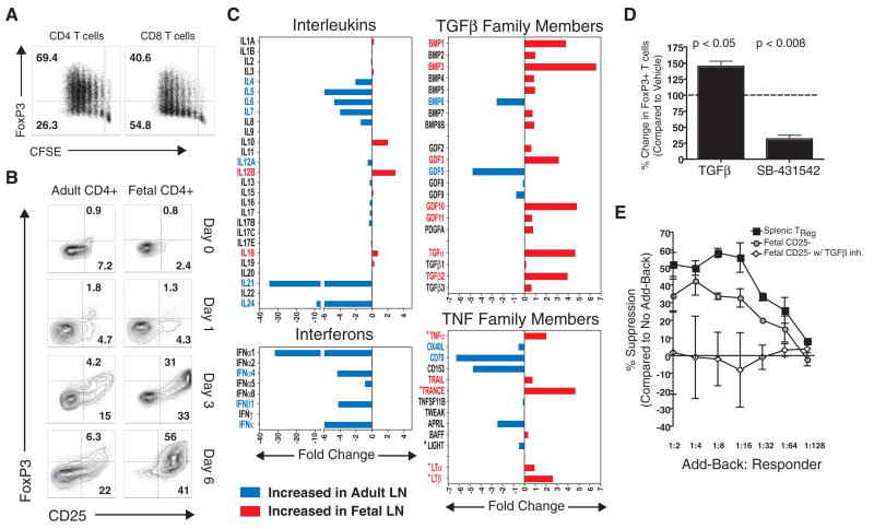
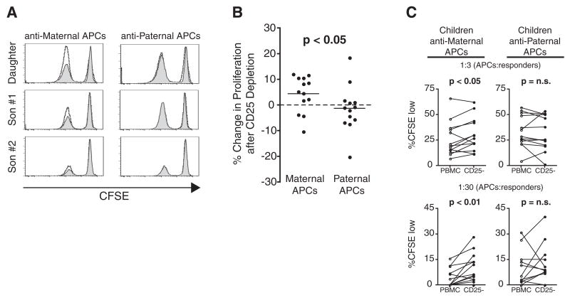
As the immune system develops, T cells are selected or regulated to become tolerant of self antigens and reactive against foreign antigens. In mice, the induction of such tolerance is thought to be attributable to the deletion of self-reactive cells. Here, we show that the human fetal immune system takes advantage of an additional mechanism: the generation of regulatory T cells (Tregs) that suppress fetal immune responses. We find that substantial numbers of maternal cells cross the placenta to reside in fetal lymph nodes, inducing the development of CD4+CD25highFoxP3+ Tregs that suppress fetal antimaternal immunity and persist at least until early adulthood. These findings reveal a form of antigen-specific tolerance in humans, induced in utero and probably active in regulating immune responses after birth.


Figures

Fig. 1
Fig. 1
Fetal Tregs suppress fetal T cell responses to maternal alloantigens. (A) Fetal T cell proliferation after stimulation with allogeneic APCs from an unrelated donor for 5 days (3:1 ratio of fetal lymphocytes:allogeneic APCs). (B) Proliferative responses to autologous, maternal, or unrelated APCs after a 5-day MLR. Histograms depict proliferation in the presence (no depletion) or absence (CD25 depletion) of fetal Tregs. CFSE, carboxy-fluorescein diacetate succinimidyl ester. (C) Summary of all experiments addressing fetal T cell proliferative responses to autologous (n = 6), maternal (n = 9), or unrelated (n = 9) APCs in the presence or absence of fetal Tregs. Statistical significance was determined by paired Student’s t test. mLN, mesenteric lymph nodes. (D) Comparison of Tregs suppression against autologous, maternal, or unrelated APCs.
Fig. 2
Fig. 2
Fetal T cells differentiate into Tregs upon stimulation with alloantigens. (A) Fetal T cells depleted of CD25+FoxP3+ cells were stimulated for 5 days with unrelated APCs, and FoxP3 expression was measured in proliferating T cells. (B) Kinetic analysis of CD25 and FoxP3 up-regulation by adult and fetal CD4+ T cells after stimulation with alloantigens. (C) Fold difference in cytokine mRNA expression in normal adult (n = 4) and fetal (n = 5) LNs. Genes found to be significantly different are labeled in red (fetal) or blue (adult) (P < 0.05, unpaired Student’s t test). Asterisks denote TNF family members involved in organogenesis. (D) Inhibition of TGFβ signaling by addition of the activin receptor–like kinase inhibitor, SB-431542 (1 uM), which blocks FoxP3 up-regulation by fetal T cells stimulated with unrelated APCs. (E) TGFβ-dependent acquisition of suppressive function after stimulation of fetal T cells with alloantigens. Error bars indicate SD observed in three separate experiments.
Fig. 3
Fig. 3
Tregs specific for non-inherited maternal alloantigens persist long after birth. (A) CD8+ T cell proliferation in mock-depleted (gray histograms) and CD25-depleted (unshaded histograms) T cells after an 8-day MLR with maternal (left) and paternal (right) APCs. Three children from a single family are represented. (B) Summary of all children tested, comparing the relative increase in proliferation after depletion of CD25+ cells from MLRs against maternal (left) or paternal (right) APCs (1:3 ratio of APCs: responders). Statistical analysis calculated by Mann-Whitney U rank sum test. (C) Summary of individual responses of all children tested, showing proliferation of mock-depleted (PBMC) or CD25-depleted (CD25−) T cells in response to maternal or paternal APCs. Two different dilutions of APCs:responders (1:3, top; 1:30, bottom) are depicted. Statistical significance was determined by paired Student’s t test.

Comment in

References

    1. Billingham RE, Brent L, Medawar PB. Nature. 1953;172:603. - PubMed
    1. Gitlin D, Kumate J, Urrusti J, Morales C. J Clin Invest. 1964;43:1938. - PMC - PubMed
    1. Remington JS, Klein JO, Wilson CB, Baker Carol J. Infectious Diseases of the Fetus and Newborn Infant. 6. Elsevier Saunders; Philadelphia, PA: 2006. pp. 11–16.
    1. Adams KM, Nelson JL. JAMA. 2004;291:1127. - PubMed
    1. Silverstein AM. Science. 1964;144:1423. - PubMed

Publication types

MeSH terms