. 2008 Dec 9;105(49):19426-31.
doi: 10.1073/pnas.0805230105. Epub 2008 Dec 4.

Stabilization of HIF-1alpha is critical to improve wound healing in diabetic mice

Affiliations

Stabilization of HIF-1alpha is critical to improve wound healing in diabetic mice

Ileana Ruxandra Botusan et al. Proc Natl Acad Sci U S A. .

Abstract

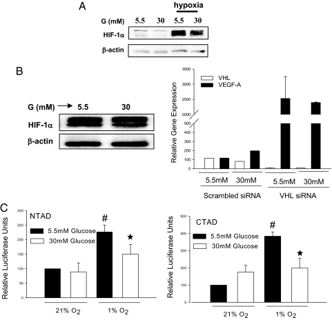
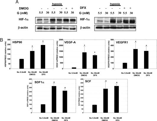
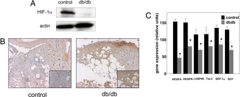
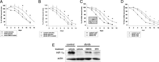
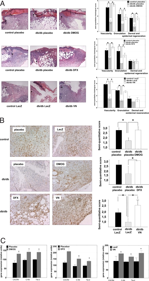
Relative hypoxia is essential in wound healing since it normally plays a pivotal role in regulation of all the critical processes involved in tissue repair. Hypoxia-inducible factor (HIF) 1alpha is the critical transcription factor that regulates adaptive responses to hypoxia. HIF-1alpha stability and function is regulated by oxygen-dependent soluble hydroxylases targeting critical proline and asparaginyl residues. Here we show that hyperglycemia complexly affects both HIF-1alpha stability and activation, resulting in suppression of expression of HIF-1 target genes essential for wound healing both in vitro and in vivo. However, by blocking HIF-1alpha hydroxylation through chemical inhibition, it is possible to reverse this negative effect of hyperglycemia and to improve the wound healing process (i.e., granulation, vascularization, epidermal regeneration, and recruitment of endothelial precursors). Local adenovirus-mediated transfer of two stable HIF constructs demonstrated that stabilization of HIF-1alpha is necessary and sufficient for promoting wound healing in a diabetic environment. Our findings outline the necessity to develop specific hydroxylase inhibitors as therapeutic agents for chronic diabetes wounds. In conclusion, we demonstrate that impaired regulation of HIF-1alpha is essential for the development of diabetic wounds, and we provide evidence that stabilization of HIF-1alpha is critical to reverse the pathological process.


Conflict of interest statement

The authors declare no conflict of interest.

Figures

Fig. 1.
Fig. 1.
Hyperglycemia-dependent destabilization and inhibition of HIF-1α. (A) Immunoblot detecting HIF-1α in db/db mouse primary fibroblasts cultured for 48 h in different glucose concentrations (5.5 mM and 30 mM) and then exposed for 6 h in either normoxia (21% O2) or hypoxia (1% O2). (B Left) immunoblot detecting HIF-1α in renal carcinoma cells (SKRC7) expressing functionally inactive VHL cells (SKRC7). B (Right) Relative expression of VEGF in human dermal fibroblast cells exposed to different glucose concentrations after transfection with VHL-specific siRNA or scrambled siRNA. (C) Relative luciferase activity in the extract of 3T3 cells exposed to different oxygen and glucose concentrations after co-transfection with CTAD (GAL4/mHIF-1α 772–822) or NTAD (GAL4/mHIF1α 531–584) and GAL4-responsive reporter gene plasmid (*, P < 0.05, 5.5 mM glucose vs. 30 mM glucose).
Fig. 2.
Fig. 2.
Inhibition of HIF hydroxylases can reverse the glucose inhibition of HIF-1α. (A) Immunoblots detecting HIF-1α in mouse db/db skin fibroblasts show destabilization of HIF-1α in high glucose concentrations (30 mM). This effect is overcome by treatment with hydroxylase inhibitors (DMOG [2 mM] or DFX [100 μM]). (B) Hydroxylase inhibitors induce expression of HIF-1α target genes essential for wound healing (DMOG [2 mM] or DFX [100 μM]) even in the presence of high glucose levels (*, P < 0.05, treatment vs. control). (No, normoxia (21% O2); Hy, hypoxia (1% O2); G, glucose.)
Fig. 3.
Fig. 3.
HIF-1α function is negatively regulated in diabetic wounds. (A) Wounds in diabetic mice are more hypoxic than in normoglycemic control mice as evaluated by pimonidazole adduct formation; note the detailed granulation tissue (Inset). (B) Immunoblot detecting HIF-1α in a whole-cell extract from the wound of diabetic or normoglycemic mice. (C) The expression of HIF target genes involved in wound healing is down-regulated in wounds of db/db mice (*, P < 0.05 expression in db/db mice vs. normoglycemic litter-mates).
Fig. 4.
Fig. 4.
Local stabilization and activation of HIF-1α by hydroxylase inhibitors or direct transfer of stabilized HIF improve wound healing in diabetic mice. (Top) The healing rate of full-thickness wounds in db/db mice is promoted by local treatment with DMOG (2 mM; A1), DFX (1 mM; B1) or by adenovirus-transferred of stable forms of HIF-1α (V-N; C1), (V-NC; D1) compared with placebo or empty virus (LacZ), respectively (values are means ± SEM; *P < 0.05 db/db treated vs. db/db placebo or LacZ). C1, Inset: virus expression at the edge of the wound as revealed by β-galactosidase staining. (Lower) Representative examples showing wound healing in db/db mice treated as in Upper. (D). Immunoblot detecting HIF-1α in whole-cell protein extracts from wounds. Note that inhibition of hydroxylase activity by local treatment with either DMOG (2 mM) or DFX (1 mM) overcomes hyperglycemia-dependent negative regulation of HIF-1α.
Fig. 5.
Fig. 5.
Stabilization and activation of HIF-1α in diabetic wounds is followed by activation of granulation angiogenesis and recruitment of CAG. (A) Hematoxylin and eosin staining shows improvement in the granulation tissue and visualization of the wounds in db/db mice locally treated with DMOG (2 mM) or DFX (1 mM) or by adenovirus-mediated expression of a stable HIF-1α (V-N; original magnification ×25). (Right) Semiquantitative evaluation for granulation, angiogenesis, and dermal and epidermal regeneration. Graphs represent means ± SEM. Vehicle-treated or empty virus treated (LacZ) normoglycemic heterozygous db mice (black bars), vehicle-treated or empty virus treated (LacZ) homozygous diabetic db/db mice (gray bars), and DMOG, DFX, or HIF V-N treated homozygous db mice (white bars) are shown (*, P < 0.05). (B) Left: vascular density in wounds evaluated by Griffonia simplicifolia 1 (GS-1) lectin staining is increased in the db/db diabetic animals after treatment with DMOG (2 mM), DFX (1 mM), or V-N. (Right) The semiquantitative evaluation of vessel density evaluated by GS-1 lectin staining. Vehicle-treated or LacZ-treated heterozygous normoglycemic mice (black bars), vehicle-treated or Lac Z-treated homozygous diabetic mice (gray bars), and DMOG, DFX, or V-N-treated homozygous diabetic mice (white bars) are shown. Graphs represent mean ± SEM. (*, P < 0.05). (C) Local treatment with DMOG, DFX, or V-N in wounds of diabetic mice increases mRNA expression of cytokine receptors typically present on CAG (*, P < 0.05 treated vs. placebo or LacZ).

References

    1. Boulton AJ, Vileikyte L, Ragnarson-Tennvall G, Apelqvist J. The global burden of diabetic foot disease. Lancet. 2005;366:1719–1724. - PubMed
    1. Dinh TL, Veves A. Treatment of diabetic ulcers. Dermatol Ther. 2006;19:348–355. - PubMed
    1. Edmonds ME. The diabetic foot, 2003. Diabetes Metab Res Rev. 2004;20(suppl 1):S9–S12. - PubMed
    1. Tandara AA, Mustoe TA. Oxygen in wound healing–more than a nutrient. World J Surg. 2004;28:294–300. - PubMed
    1. Knighton DR, Silver IA, Hunt TK. Regulation of wound-healing angiogenesis-effect of oxygen gradients and inspired oxygen concentration. Surgery. 1981;90:262–270. - PubMed

Publication types

MeSH terms