Review
. 2009 Jul;29(7):992-1000.
doi: 10.1161/ATVBAHA.108.165506. Epub 2008 Dec 4.

Nanotechnology in medical imaging: probe design and applications

Affiliations
Review

Nanotechnology in medical imaging: probe design and applications

David P Cormode et al. Arterioscler Thromb Vasc Biol. 2009 Jul.

Abstract

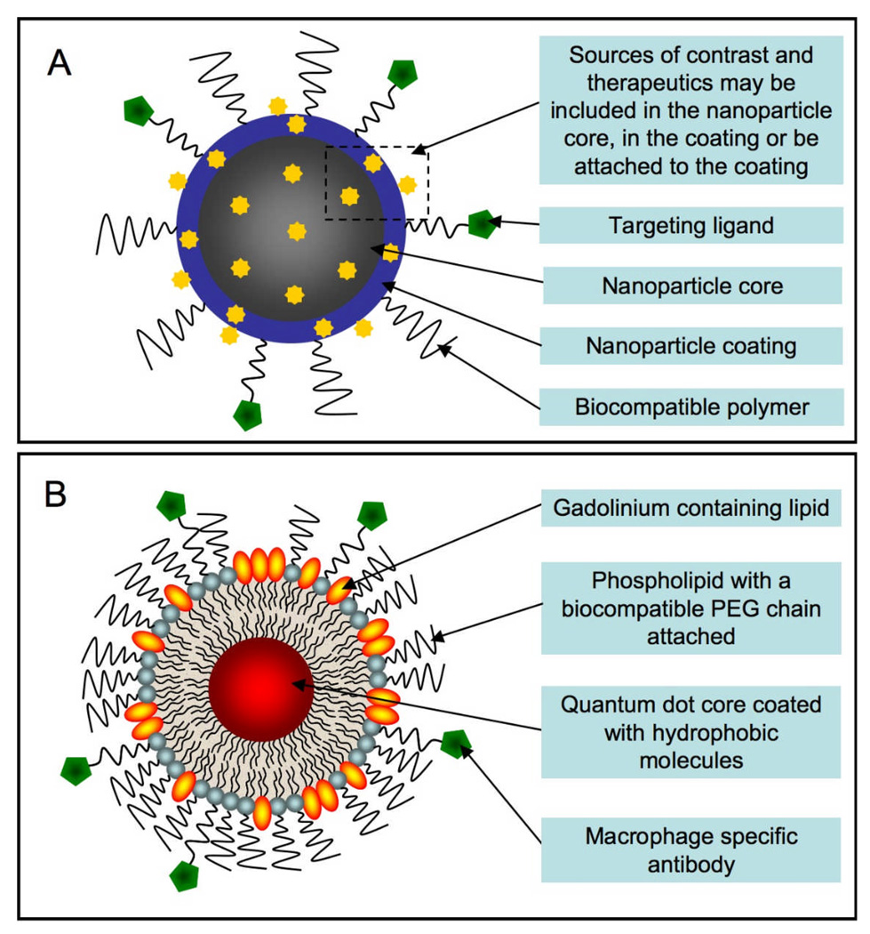
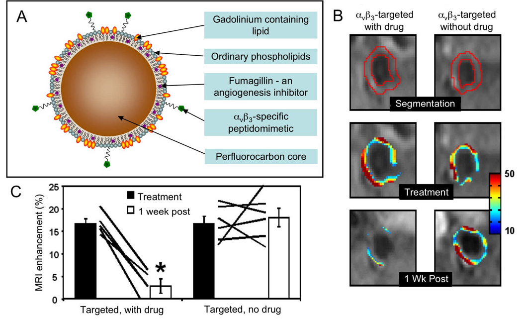
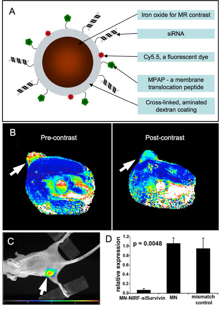
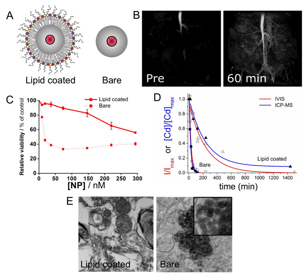
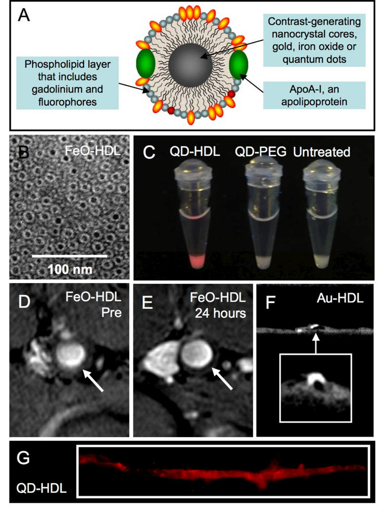
Nanoparticles have become more and more prevalent in reports of novel contrast agents, especially for molecular imaging, the detection of cellular processes. The advantages of nanoparticles include their potency to generate contrast, the ease of integrating multiple properties, lengthy circulation times, and the possibility to include high payloads. As the chemistry of nanoparticles has improved over the past years, more sophisticated examples of nano-sized contrast agents have been reported, such as paramagnetic, macrophage targeted quantum dots or alpha(v)beta(3)-targeted, MRI visible microemulsions that also carry a drug to suppress angiogenesis. The use of these particles is producing greater knowledge of disease processes and the effects of therapy. Along with their excellent properties, nanoparticles may produce significant toxicity, which must be minimized for (clinical) application. In this review we discuss the different factors that are considered when designing a nanoparticle probe and highlight some of the most advanced examples.


Figures

Figure 1
Figure 1
A Generalized schematic of a nanoparticle-based molecular imaging contrast agent. B Schematic of a quantum dot based paramagnetic contrast agent targeted to macrophages via CD204 antibodies.
Figure 2
Figure 2
A schematic depiction of a microemulsion based contrast agent/drug delivery nanoparticle. B MR images of the aortas of hyperlipidemic rabbits injected with the microemulsion contrast agent with or without the anti-angiogenesis drug fumagillin. The superimposed color maps denote the percent signal enhancement in the aorta. C quantification of enhancement upon agent injection pre and one week post-treatment showing reduction of angiogenesis when the drug was included in the particle. Parts of this figure reproduced, with permission, from reference .
Figure 3
Figure 3
A Schematic depiction of an iron oxide based MR contrast agent that can deliver siRNA. B MR image of a mouse pre- and 24 hrs post-administration of the agent. Change in pixel color in the tumor (arrow) from red-yellow to blue indicates accumulation of the agent. C Fluorescence image of the mouse with emission seen from the tumor (arrow), also indicating agent accumulation. D Silencing of the Birc5 gene by the siRNA labeled nanoparticle (MN-NIRF-siSurvivin) reduces Survivin expression in the tumor and leads to apoptosis in this tissue. Parts of this figure reproduced, with permission, from reference .
Figure 4
Figure 4
A Schematic depictions of lipid-coated and bare silica particles that have a quantum dot in their cores. B MRI of the vasculature of a mouse before and after administration of the paramagnetic, PEG-lipid coated silica particles. C Cell viability assays indicating lowered cytotoxicity of the lipid-coated particles. D Blood clearance profiles of the silica particles showing the enhanced circulation half-life of the coated particles. E Transmission electron micrographs of mouse lungs after injection with silica particles. Large aggregates of the particles were found in the lungs of mice injected with bare particles. The images in this figure are reproduced, with permission, from reference .
Figure 5
Figure 5
A Schematic depiction of nanocrystal-core high density lipoprotein (HDL). B Negative stain TEM of FeO-HDL. C Photograph of pellets of cells incubated with QD-HDL, QD-PEG or without a contrast agent taken under UV irradiation. MR images of the aorta of an apoE KO mouse taken D before and E 24 hours after injection of FeO-HDL. F Micro-CT image of an excised aorta of a mouse injected 24 hours previously with Au-HDL. G Fluorescence image of an excised aorta of a mouse injected 24 hours previously with QD-HDL. Parts of this figure reproduced, with permission, from reference .

References

    1. Corot C, Robert P, Idee J-M, Port M. Recent advances in iron oxide nanocrystal technology for medical imaging. Adv. Drug Deliv. Rev. 2006;58:1471–1504. - PubMed
    1. Mulder WJM, Griffioen AW, Strijkers GJ, Cormode DP, Nicolay K, Fayad ZA. Magnetic and fluorescent nanoparticles for multimodality imaging. Nanomedicine. 2007;2:307–324. - PubMed
    1. Huang X, Jain PK, El-Sayed IH, El-Sayed M. Gold nanoparticles: interesting optical properties and recent applications in cancer diagnostics and therapy. Nanomedicine. 2007;2:681–693. - PubMed
    1. Azzazy HME, Mansour MMH, Kazmierczak SC. From diagnostics to therapy: Prospects of quantum dots. Clin. Biochem. 2007;40:917–927. - PubMed
    1. Wickline SA, Neubauer AM, Winter PM, Caruthers SD, Lanza GM. Molecular imaging and therapy of atherosclerosis with targeted nanoparticles. J. Magn. Reson. Imaging. 2007;25:667–680. - PubMed

Publication types