. 2009 Jan 7;28(1):21-33.
doi: 10.1038/emboj.2008.238. Epub 2008 Dec 4.

PTEN regulation by Akt-EGR1-ARF-PTEN axis

Affiliations

PTEN regulation by Akt-EGR1-ARF-PTEN axis

Jianxiu Yu et al. EMBO J. .

Abstract

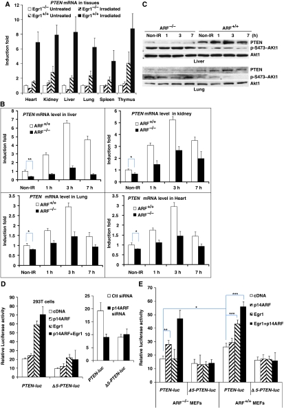
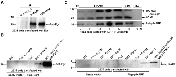
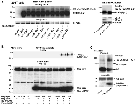
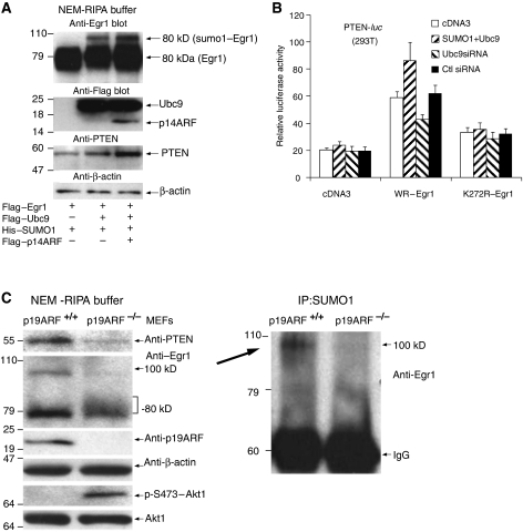
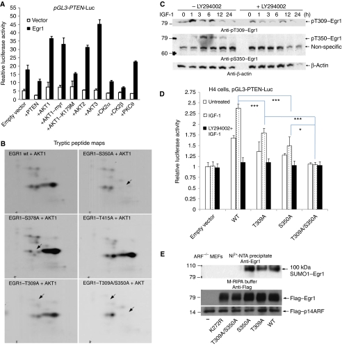
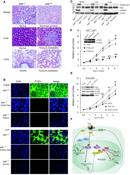
The PTEN tumour suppressor gene is induced by the early growth response 1 (EGR1) transcription factor, which also transactivates p53, p73, and p300/CBP as well as other proapoptotic and anti-cancer genes. Here, we describe a novel Akt-EGR1-alternate reading frame (ARF)-PTEN axis, in which PTEN activation in vivo requires p14ARF-mediated sumoylation of EGR1. This modification is dependent on the phosphorylation of EGR1 at S350 and T309 by Akt, which promotes interaction of EGR1 with ARF at K272 in its repressor domain by the ARF/Ubc9/SUMO system. EGR1 sumoylation is decreased by ARF reduction, and no EGR1 sumoylation is detected in ARF(-/-) mice, which also exhibit reduced amounts of PTEN. Our model predicts that perturbation of any of the clinically important tumour suppressors, PTEN, EGR1, and ARF, will cause some degree of dysfunction of the others. These results also explain the known negative feedback regulation by PTEN on its own synthesis through PI3 kinase inhibition.


Figures

Figure 1
Figure 1
PTEN is not induced in tissues of the EGR1−/− or ARF−/− mouse models. (A) Analysis of PTEN mRNA levels by qRT–PCR in six tissues from 8-week-old, healthy EGR1−/− and wild-type mice that were γ-irradiated with 5 Gy and killed 2.5 h later. (B) A similar qRT–PCR analysis for PTEN mRNA in four tissues: liver, kidney, lung, and heart (and also spleen, data not shown) from ARF−/− 129sv/BL6 mice and wild-type mice of the same strain that were γ-irradiated with5 Gy and killed 0, 1, 3, or 7 h later. (C) Western blot analysis for PTEN, Akt1, and phospho-Akt1 in two tissues liver and lung from the same mice in (B). Luciferase activity measured 24 h after transfection into 293T cells (D) ARF−/− and ARF+/+ MEFs (E) with the indicated DNA constructs and siRNAs. The PTEN-luciferase promoter construct was described earlier (Virolle et al, 2001) and the same reporter Δ5-PTEN-luc lacking most EGR1-binding sites is shown in Supplementary Figure S2. The data represent the mean and triplicate determinations±s.d. (*P<0.05, **P<0.01, ***P<0.001).
Figure 2
Figure 2
EGR1 interacts with ARF. (A) Anti-EGR1 immunoblot of normal IgG (control) or anti-p14ARF immunoprecipitates from 293T cells transfected with EGR1. Lane 3 contains input cell lysate (in M-RIPA buffer). (B) Left panel: equal amounts of bacterially expressed GST or GST–p14ARF fusion proteins and Glutathione-Sepharose beads were incubated with lysates of 293T cells transfected with empty vector or vector-EGR1. The proteins associated with GST–p14ARF, bound on the Glutathione-Sepharose beads, were washed five times with the same M-RIPA buffer before western blotting. The input is the cell lysate of cells transfected with EGR1. Right panel: anti-p14ARF immunoblot of material bound to the indicated GST or GST–EGR1 fragments N(1–278), M(274–421), or C(416–543) fusion proteins in lysates (in M-RIPA buffer) from 293T cells transfected with empty vector or Flag–p14ARF. (C) Anti-EGR1 and p14ARF immunoblot of anti-p14ARF or EGR1 or IgG immunoprecipitates from HeLa cells treated with IGF-1 for the indicated times 0–12 h (with NEM-RIPA buffer).
Figure 3
Figure 3
ARF promotes the sumoylation of EGR1. (A) Left panel: 293T cells cotransfected with wild-type or mutated EGR1 constructs with or without Ubc9/SUMO1 were directly lysed in NEM-RIPA buffer 48 h after transfection. Right panel: 293T cells cotransfected with Ubc9/SUMO1/EGR1 and control siRNA or p14ARF siRNA were lysed in NEM-RIPA buffer 48 h after transfection. Western blot was analysed with indicated antibodies. (B) Anti-EGR1 blot of Ni2+-NTA precipitates from ARF−/− MEFs (passage 10) transfected with Flag–EGR1 (K272R, K5R, and wild type) alone and with Flag–UBC9/His6–SUMO1 and with or without Flag–p14ARF, as indicated (upper panel); anti-Flag blot of total lysates in NEM-RIPA buffer (lower panel). (C) Anti-EGR1 blot of anti-SUMO1 immunoprecipitates from HeLa cells transfected with empty vector or Flag–p14ARF (upper panel). Lower panel: anti-EGR1 or anti-Flag immunoblot of total lysates.
Figure 4
Figure 4
Sumoylation of EGR1 is required for PTEN induction. (A) Anti-EGR1 blot of 293T cells transfected with Flag–EGR1 alone or with Ubc9/SUMO1 and p14ARF as indicated (upper panel). Anti-Flag (second panel), anti-PTEN (third panel), and anti-β-actin (bottom panel) blots of the same experiment. (B) Activation of the PTEN-luc reporter by WT–EGR1 and by K272R–EGR1 in the presence of Ubc9/SUMO1 or Ubc9 siRNA. The 293T cells were transfected with PTEN-luc and SUMO1/Ubc9 or control siRNA or Ubc9 siRNA, and empty vector cDNA3, or WT-EGR1, or K272R-EGR1, as indicated. Luciferase activity was measured 24 h later. (C) Left panel: immunoblots for PTEN, EGR1, p19ARF, β-actin, Akt1, and phospho-Akt1 from ARF−/− (passage 12) and ARF+/+ (passage 5) MEFs. Right panel: anti-EGR1 blot of anti-SUMO1 immunoprecipitates from the same lysates.
Figure 5
Figure 5
PTEN transactivation by Akt–EGR1–ARF–PTEN axis. (A) Left panels: EGR1, ARF, and PTEN mRNA levels assessed by qRT–PCR from HeLa cells treated for the indicated times with 100 ng/ml of IGF-1 (black symbols) or LY294002 plus IGF-1 (open symbols). Right panels: immunoblots for phospho-Akt, EGR1, EGR1 in anti-SUMO immunoprecipitates, ARF, PTEN, and β-actin from the same IGF-1-treated cells. The second panel in each group is from cells treated with 20 μM of the PI3K inhibitor LY294002. SUMO1 immunoprecipitations were performed in the presence of N-ethylmaleimide to block desumoylation. (B) Immunoblots of lysates from ARF+/+ and ARF−/− embryonic fibroblasts treated as in (A). (C) EGR1 and p14ARF location as a function of time after addition of IGF-1. Confocal microscopy of EGR1 (red), p14ARF (green), and DNA (blue) in cells treated with IGF-1 for the indicated times. Note that some EGR1s and ARFs colocalize at peak between 3 and 6 h. These confocal images are not taken with equal exposure times and therefore protein levels cannot be compared between panels, for example, it does not show here that EGR1 protein levels are much lower at t=0 than at t=1 h. The purpose of this experiment was to visualize the location of EGR1 and ARF, rather than quantitate. The lower row of images are parallel samples treated with the PI3K inhibitor LY294002 where p14ARF is much reduced. All individual and merged stains are shown in Supplementary Figure S7.
Figure 6
Figure 6
Akt-phosphorylated EGR1 is necessary for EGR1 sumoylation and PTEN induction. (A) Transactivation of the PTEN-luc reporter by EGR1 in the presence of PTEN, Akt, or other protein kinases. The pGL3–PTEN–luc construct was transfected into 293T cells with empty pcDNA3 vector or EGR1 in the same vector together with expression plasmids for PTEN or the indicated kinases. Luciferase activity was measured 24 h later. Note that PTEN decreases PTEN transcription, whereas all three forms of Akt increase PTEN transcription. Akt1–K179M is a kinase-inactive mutant of Akt1, whereas Akt1myr is constitutively active. All kinases were well expressed (not shown). (B) Tryptic peptide maps of the indicated mutant EGR1-M proteins phosphorylated by Akt1. Identification of S350 and T309 as phosphorylation sites in the Zn-finger region of EGR1. (C) Phosphorylations of EGR1 at S350 and T309 in cells. Lysates from HeLa cell samples in parallel as Figure 5A were blotted with anti-pS350 or anti-pT309 or anti-β-actin. (D) Transactivation of the PTEN-luc reporter by point-mutated EGR1. The PTEN-luc construct was transfected into H4 cells together with empty vector or with the wild type or the indicated point-mutants of EGR1. Twenty-four hours after transfection, serum-free medium was used for 18 h, and to one group LY294002 (20 μM) was added for 1 h pretreatment. Luciferase activity was measured 12 h later after addition of IGF-1 (100 ng/ml). (E) EGR1 phosphorylation at two sites T309 and S350 by Akt is necessary for EGR1 sumoylation. Anti-EGR1 blot of Ni2+-NTA precipitates from ARF−/− MEFs (passage 10) transfected with UBC9/His6–SUMO1 and Flag–p14ARF together with (or without) indicated mutants or wild-type Flag–EGR1. Anti-Flag blot of total lysates (lower two panels) in M-RIPA buffer. This experiment is similar to Figure 3B.
Figure 7
Figure 7
No SUMO1–EGR1 and low expression of PTEN in ARF−/− mouse tissues. (A) Upper panel: sections from an ARF−/− mouse (8 months old) with poorly differentiated sarcoma (primary tumours were in kidney) revealing fascicles of spindle cells, compared with sections from an ARF+/+ mouse normal kidney tissues. Middle panel: sections from the same ARF−/− mouse reveal liver metastasis (arrows) from sarcoma, compared with sections from ARF+/+ mouse liver tissues. Lower panel: sections from the same ARF−/− mouse reveal lung metastasis from the sarcoma, compared with sections from lung tissues of the ARF+/+ mouse. (B) Sections (normal and tumorous) from the same ARF−/−, ARF+/+ tissues as in Figure 7A were immunostained with antibody to PTEN (green) and DAPI (blue). (C) Immunoblot of anti-EGR1 in SUMO1 immunoprecipitates (upper panel), and immunoblot of anti-EGR1 (second panel), anti-p19ARF (third panel) and anti-β-actin (bottom panel) in the same tissue lysates. Note that sumoylated EGR1 is only highly expressed in ARF+/+ mouse tissues. New pathway suppresses cell proliferation by control of PTEN expression. ARF−/− MEFs infected with pBabe–PuroL–PTEN (WT), pBabe–PuroL–PTEN (C124S), pBabe–PuroL vector (D) and wild-type primary MEFs infected with pBabe–PuroL–EGR1 (WT), pBabe–PuroL–EGR1 (K272R), pBabe–PuroL vector (E) were seeded on plates at 4–5 × 10³ cells in 96-well plates and cell numbers were determined by CyQUANT fluorescence assay at 0.5, 1, 2, 3, 4, 5 days after seeding. The cell numbers were normalized against the value at 0.5 day (Mean values±s.e.m., from three independent experiments, *P<0.05, **P<0.01, ***P<0.001). The related protein levels were shown by western blot (inset). (F) Schematic model of the PTEN transactivation pathway through a novel Akt–EGR1–ARF–PTEN axis.

References

    1. Birle D, Bottini N, Williams S, Huynh H, deBelle I, Adamson E, Mustelin T (2002) Negative feedback regulation of the tumor suppressor PTEN by phosphoinositide-induced serine phosphorylation. J Immunol 169: 286–291 - PubMed
    1. Carrasco DR, Fenton T, Sukhdeo K, Protopopova M, Enos M, You MJ, Divicio D, Nogueira C, Stommel J, Pinkus GS, Fletcher C, Hornick JL, Cavenee WK, Furnari FB, DePinho RA (2006) The PTEN and INK4A/ARF tumor suppressors maintain myelolymphoid homeostasis and cooperate to constrain histiocytic sarcoma development in humans. Cancer Cell 9: 379–390 - PubMed
    1. Chen L, Chen J (2003) MDM2–ARF complex regulates p53 sumoylation. Oncogene 22: 5348–5357 - PubMed
    1. Davies MA, Koul D, Dhesi H, Berman R, McDonnell TJ, McConkey D, Yung WKA, Steck PA (1999) Regulation of Akt/PKB activity, cellular growth, and apoptosis in prostate carcinoma cells by MMAC/PTEN. Cancer Res 59: 2551–2556 - PubMed
    1. de Belle I, Huang RP, Fan Y, Liu C, Mercola D, Adamson ED (1999) p53 and Egr-1 additively suppress transformed growth in HT1080 cells but Egr-1 counteracts p53-dependent apoptosis. Oncogene 18: 3633–3642 - PubMed

Publication types