. 2008:14:2227-36.
Epub 2008 Dec 8.

Double homozygous waltzer and Ames waltzer mice provide no evidence of retinal degeneration

Affiliations

Double homozygous waltzer and Ames waltzer mice provide no evidence of retinal degeneration

Zubair M Ahmed et al. Mol Vis. 2008.

Abstract

Purpose: To determine whether cadherin 23 and protocadherin 15 can substitute for one another in the maintenance of the retina and other tissues in the mouse. Does homozygosity for both v and av mutant alleles (i.e., a double homozygous mouse) cause retinal degeneration or an obvious retinal histopathology?

Methods: We generated mice homozygous for both Cdh23(v-6J) and Pcdh15(av-Jfb) alleles. The retinal phenotypes of double heterozygous and double homozygous mutant mice were determined by light microscopy and electroretinography (ERG). Histology on 32 different tissues, scanning electron microscopy of organ of Corti hair cells as well as serum biochemical and hematological examinations were evaluated.

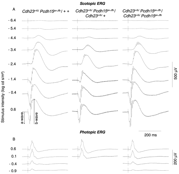
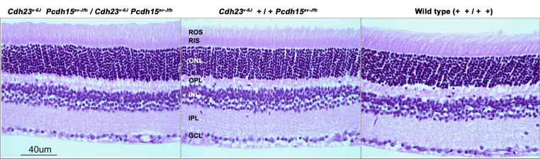
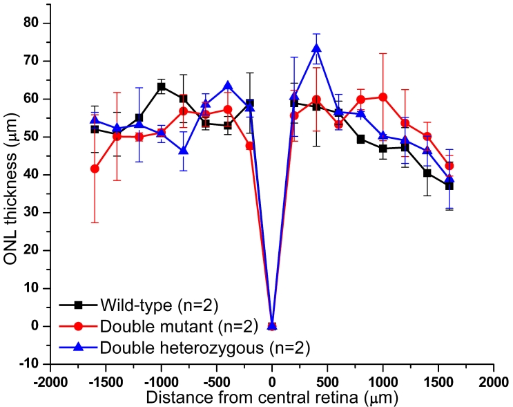
Results: ERG waves of double heterozygous and double homozygous mice showed similar shape, growth of the amplitude with intensity, and implicit time for both rod and cone pathway mediated responses. Mice homozygous for both Cdh23(v-6J) and Pcdh15(av-Jfb) mutations showed no sign of retinitis pigmentosa or photoreceptor degeneration but, as expected, were deaf and had disorganized hair cell sensory bundles.

Conclusions: The simultaneous presence of homozygous mutant alleles of cadherin 23 and protocadherin 15 results only in deafness, not retinal degeneration or any other additional obvious phenotype of the major organ systems. We conclude that in the mouse cadherin 23 or protocadherin 15 appear not to compensate for one another to maintain the retina.


Figures

Figure 1
Figure 1
Electroretinogram waveforms recorded from 5-month-old mice. A: Double heterozygous mouse (+ +/Cdh23v-6J Pcdh15av-Jfb), a Cdh23v-6J homozygote that is heterozygous for the Pcdh15av-Jfb allele (Cdh23v-6J +/Cdh23v-6J Pcdh15av-Jfb), and a double homozygous (Cdh23v-6J Pcdh15av-Jfb/Cdh23v-6J Pcdh15av-Jfb) mouse. Dark-adapted scotopic responses to the stimulus intensity range from −6.9 to +0.6 log cd-s/m2. B: Light-adapted photopic responses to the stimulus intensity range from −0.9 to +0.6 log cd-s/m2 on a 34 cd/m2 background.
Figure 2
Figure 2
Light photomicrographs of retinal morphology of a wild-type, a double heterozygous (+ +/Cdh23v-6J Pcdh15av-Jfb), and a double homozygous (Cdh23v-6J Pcdh15av-Jfb/Cdh23v-6J Pcdh15av-Jfb) mouse at P120. No structural abnormalities in the retinas of these mice were observed when compared to the wild-type mice. Images are of the mid-peripheral retina in sections taken close to the optic nerve of each eye. Abbreviations: rod outer segment (ROS); rod inner segments (RIS); outer nuclear layer (ONL); outer plexiform layer (OPL); inner nuclear layer (INL); inner plexiform layer (IPL); ganglion cell layer (GCL).
Figure 3
Figure 3
Measurements of outer nuclear layer in wild-type, heterozygous (+ +/Cdh23v-6J Pcdh15av-Jfb), and double homozygous (Cdh23v-6J Pcdh15av-Jfb/Cdh23v-6J Pcdh15av-Jfb) mice. Measurements were performed every 200 µm from central retina close to the optic nerve head to an area near the peripheral edge of retinal sections. One-way ANOVA of mean outer nuclear layer (ONL) width measurements indicate no significant effect of double homozygosity for av and v mutant alleles.
Figure 4
Figure 4
Scanning electron micrographs of the outer hair cells from the middle cochlear turn of A: P2 Cdh23v-6J heterozygote (+ +/Cdh23v-6J +), B: Cdh23v-6J +/Cdh23v-6J Pcdh15av-Jfb mouse, C: + Pcdh15av-Jfb/Cdh23v-6J Pcdh15av-Jfb mouse, and D: Cdh23v-6J Pcdh15av-Jfb/Cdh23v-6J Pcdh15av-Jfb mouse. Inset shows a close-up view of a stereocilia bundle of a Cdh23v-6J Pcdh15av-Jfb/Cdh23v-6J Pcdh15av-Jfb mouse. Note the presence of stereocilia links. Scale bars in A-D equal 2 μm; scale bar in inset in D equals 200 nm.
Figure 5
Figure 5
Schematic representation of the Cdh23 gene structure and splice variants. Isoform a of Cdh23 is the full-length transcript and contains 69 coding (black) and noncoding (gray) exons [18]. Isoforms b and c were previously reported [29]. Exon 68 encodes part of the cytoplasmic domain of cadherin 23 and is alternatively spliced. Two alternatively splice variants of isoform a, involving exons 5–10 (GenBank EU681829 and EU681830) of Cdh23 were amplified from inner ear and retina cDNA using PCR primers located in the sequence of exons 5 and 13. Cdh23v-6J is a nonsense mutation found in exon 9, which is absent in one of the alternative splice variants of Cdh23 isoform a.

References

    1. Ahmed ZM, Riazuddin S, Wilcox ER. The molecular genetics of Usher syndrome. Clin Genet. 2003;63:431–44. - PubMed
    1. Smith RJ, Berlin CI, Hejtmancik JF, Keats BJ, Kimberling WJ, Lewis RA, Möller CG, Pelias MZ, Tranebjaerg L. Clinical diagnosis of the Usher syndromes. Usher Syndrome Consortium. Am J Med Genet. 1994;50:32–8. - PubMed
    1. Ahmed Z, Riazuddin S, Khan S, Friedman P, Riazuddin S, Friedman T. USH1H, a novel locus for type I Usher syndrome, maps to chromosome 15q22–23. Clin Genet. 2008 - PMC - PubMed
    1. Petit C. Usher syndrome: from genetics to pathogenesis. Annu Rev Genomics Hum Genet. 2001;2:271–97. - PubMed
    1. Gerber S, Bonneau D, Gilbert B, Munnich A, Dufier JL, Rozet JM, Kaplan J. USH1A: Chronicle of a Slow Death. Am J Hum Genet. 2006;78:357–9. - PMC - PubMed

Publication types

LinkOut - more resources