. 2008 Dec;4(12):e1000291.
doi: 10.1371/journal.pgen.1000291. Epub 2008 Dec 5.

Targeting the X chromosome during spermatogenesis induces Y chromosome transmission ratio distortion and early dominant embryo lethality in Anopheles gambiae

Affiliations

Targeting the X chromosome during spermatogenesis induces Y chromosome transmission ratio distortion and early dominant embryo lethality in Anopheles gambiae

Nikolai Windbichler et al. PLoS Genet. 2008 Dec.

Abstract

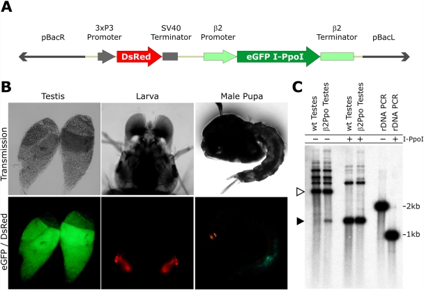
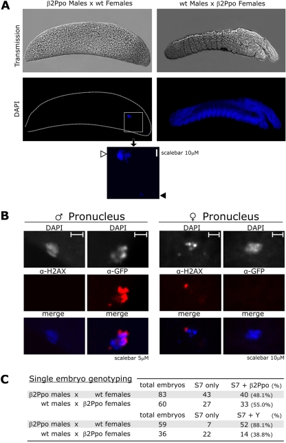
We have exploited the high selectivity of the homing endonuclease I-PpoI for the X-linked Anopheles gambiae 28S ribosomal genes to selectively target X chromosome carrying spermatozoa. Our data demonstrated that in heterozygous males, the expression of I-PpoI in the testes induced a strong bias toward Y chromosome-carrying spermatozoa. Notably, these male mosquitoes also induced complete early dominant embryo lethality in crosses with wild-type females. Morphological and molecular data indicated that all spermatozoa, irrespectively of the inheritance of the transgene, carried a substantial amount of I-PpoI protein that could attack the maternally inherited chromosome X of the embryo. Besides the obvious implications for implementing vector control measures, our data demonstrated the feasibility of generating synthetic sex distorters and revealed the intriguing possibility of manipulating maternally inherited genes using wild-type sperm cells carrying engineered endonucleases.


Conflict of interest statement

The authors have declared that no competing interests exist.

Figures

Figure 1
Figure 1. Transformation construct and expression of I-PpoI in the testes of transgenic mosquitoes.
(A) Schematic representation of the construct pBac{3xP3-DsRed}β2-eGFP::I-PpoI containing the left and right piggyBac inverted repeats (pBacR,L); the Pax promoter (3xP3) driving the DsRed marker gene; and the eGFP::I-PpoI effector gene (eGFP I-PpoI) under the control of β2 tubulin promoter and terminator (β2). (B) Transmission and fluorescent images of dissected adult testes, larval head and pupa of β2Ppo1 male mosquitoes. (C) Southern blot analysis of the 28S ribosomal DNA locus. DNA from testes of WT (lanes 1 and 3) and β2Ppo1 males (lanes 2 and 4) was digested with the endonuclease ClaI in vitro and hybridized with a probe encompassing the 28S ribosomal gene (Figure S1). As control both the DNA extracted from the WT and β2Ppo1 testes was treated with recombinant I-PpoI as indicated. Furthermore the PCR product (2kb) used as probe either treated with recombinant I-PpoI or untreated was analysed under the same hybridization conditions (lanes 4 and 5). Open and filled arrowheads indicate the full length and digested rDNA fragments respectively.
Figure 2
Figure 2. Confocal analysis of spermatozoa from β2Ppo and WT males recovered from spermathecae of WT females.
(A) Spermatheca of a female mated with β2Ppo1 males analysed by transmission microscopy (left), analyzed for eGFP fluorescence (middle) and DNA stained with DAPI (right). (B) Analysis of confocal 3D data stacks of sperm extracted from spermathecae of females mated to WT or β2Ppo males. Objects defined as sperm, on the basis of DAPI fluorescence and size, were analyzed in a way that GFP density (nuclear volume/fluorescence intensity) was plotted against DAPI density. Density values were plotted for each individual spermatozoa examined from wt (black rectangles) and transgenic (grey diamonds) males. (C) Assessment of mating capability of β2Ppo2 against WT males. Equal numbers (10) of β2Ppo and WT males were allowed to mate in the presence of 10 or 20 WT females for 6 days. The mating with WT and transgenic males was assessed by analyzing in PCR experiments the DNA extracted from the spermathecae using a first a marker revealing chromosome Y specific sequence, to provide an overall estimate of mating rate and a second marker for the I-PpoI coding sequence. PCRs experiments that failed to amplify any product were scored as non-mated. The figure shows the percentage of mated mosquitoes and the relative contribution of WT (grey) and transgenic males (black) in the mating. Shown is the combined average of 3 independent sets of experiments.
Figure 3
Figure 3. Morphological and genotype analysis of developmentally arrested embryos.
(A) Embryos originating from crosses between β2Ppo males with WT females (left) compared to crosses between WT males and β2Ppo females (right) were analyzed by fluorescence microscopy 24 hours after oviposititon. The figure shows transmission (upper panels) and fluorescence of DAPI staining DNA (lower panels) of embryos oriented with the posterior end to the left and ventral side up. The inset shows a magnified view of the small and large DAPI stained bodies found in the embryos marked with a black and a white arrow respectively. (B) Immunostaining of freshly hatched embryos using mouse anti GFP (α-GFP) or mouse anti γ-H2AX (α-H2AX) primary in combination with anti-mouse IgG Alexa-532 conjugated secondary antibodies. DAPI stained bodies identified as male pronucleus and female pronucleus are shown at 5 and 10 µM scale respectively. (C) Molecular genotyping of embryos using multiplex PCR. Embryos originating from crosses of β2Ppo males with WT females and WT males with β2Ppo females were collected at 24 hrs post deposition and their DNA was examined using nested PCR analysis to amplify Y chromosome or transgene specific sequences as well as the ribosomal gene S7 as a control. The values show the frequency of the genotypes in all embryos that tested positive for the presence of S7.

References

    1. World Health Organisation. Geneva: WHO; 2005. World Malaria Report 2005: Roll Back Malaria.
    1. Kouyate B, Sie A, Ye M, De Allegri M, Muller O. The great failure of malaria control in Africa: a district perspective from Burkina Faso. PLoS Med. 2007;4:e127. - PMC - PubMed
    1. Curtis CF, Graves PM. Methods for replacement of malaria vector populations. J Trop Med Hyg. 1988;91:43–48. - PubMed
    1. Beaty BJ. Genetic manipulation of vectors: A potential novel approach for control of vector-borne diseases. Proc Natl Acad Sci U S A. 2000;97:10295–10297. - PMC - PubMed
    1. Carlson J, Olson K, Higgs S, Beaty B. Molecular genetic manipulation of mosquito vectors. Annu Rev Entomol. 1995;40:359–388. - PubMed

Publication types

MeSH terms