. 2008 Dec;4(12):e1000295.
doi: 10.1371/journal.pgen.1000295. Epub 2008 Dec 5.

Apoptosis maintains oocyte quality in aging Caenorhabditis elegans females

Affiliations

Apoptosis maintains oocyte quality in aging Caenorhabditis elegans females

Sara Andux et al. PLoS Genet. 2008 Dec.

Abstract

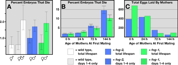
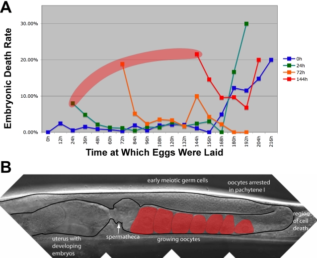
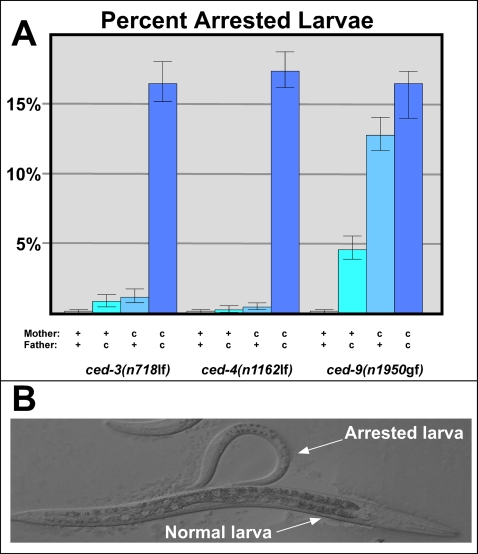
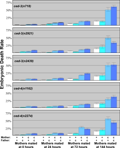
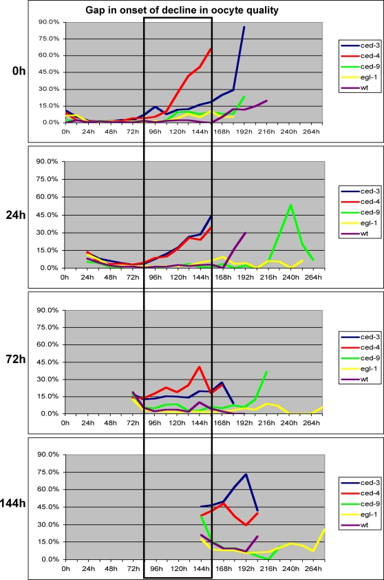
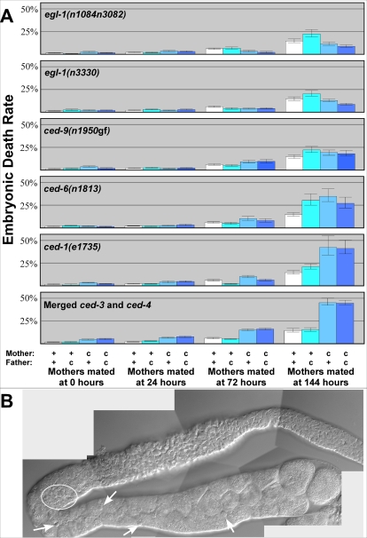
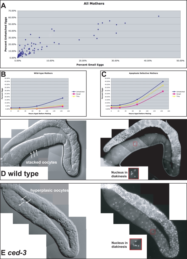
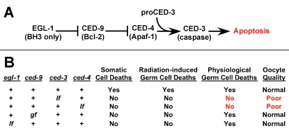
In women, oocytes arrest development at the end of prophase of meiosis I and remain quiescent for years. Over time, the quality and quantity of these oocytes decreases, resulting in fewer pregnancies and an increased occurrence of birth defects. We used the nematode Caenorhabditis elegans to study how oocyte quality is regulated during aging. To assay quality, we determine the fraction of oocytes that produce viable eggs after fertilization. Our results show that oocyte quality declines in aging nematodes, as in humans. This decline affects oocytes arrested in late prophase, waiting for a signal to mature, and also oocytes that develop later in life. Furthermore, mutations that block all cell deaths result in a severe, early decline in oocyte quality, and this effect increases with age. However, mutations that block only somatic cell deaths or DNA-damage-induced deaths do not lower oocyte quality. Two lines of evidence imply that most developmentally programmed germ cell deaths promote the proper allocation of resources among oocytes, rather than eliminate oocytes with damaged chromosomes. First, oocyte quality is lowered by mutations that do not prevent germ cell deaths but do block the engulfment and recycling of cell corpses. Second, the decrease in quality caused by apoptosis mutants is mirrored by a decrease in the size of many mature oocytes. We conclude that competition for resources is a serious problem in aging germ lines, and that apoptosis helps alleviate this problem.


Conflict of interest statement

The authors have declared that no competing interests exist.

Figures

Figure 1
Figure 1. Oocyte quality decreases with maternal age.
(A) Percent of eggs that died before hatching. The mothers were wildtype hermaphrodites (white), fog-2(q71) females (blue), and fog-1(q250) females (green). Solid bars represent the fraction of all eggs that died throughout the entire reproductive span; shaded bars represent the fraction of eggs that died during the first four days of the reproductive span. Error bars represent 95% confidence intervals. (B) Percent of eggs that died before hatching, produced by females mated at the specified ages. (C) Total eggs produced by females mated at the specified ages.
Figure 2
Figure 2. Aging affects two populations of oocytes.
(A) Percent of eggs that died before hatching, plotted against the age of the mother when the eggs were laid (in hours after the molt into adulthood). The mothers were virgin fog-2(q71) females mated at zero hours (blue), 24 hours (green), 72 hours (orange) and 144 hours (red). The shaded area indicates eggs formed from oocytes that had been arrested in diakinesis. (B) Nomarski photomicrograph of the gonad of a mated female. Arrow indicates the spermatheca. Shaded oocytes are arrested in diakinesis. Anterior is to the left, and ventral is down.
Figure 3
Figure 3. The larval lethality caused by apoptosis mutants is not due to low oocyte quality.
(A) Females with (c) or without (+) the indicated mutation were crossed with corresponding males, and their offspring were assayed for growth. Bars indicate the percent of larvae from each cross that remained in the L1 stage after 48 hours. Error bars represent 95% confidence intervals. (B) Nomarski photomicrograph of two ced-3(n718) larvae at 48 hours post-fertilization. The animal at the top had permanently arrested at this stage, and eventually died.
Figure 4
Figure 4. Blocking germ cell death lowers oocyte quality in aging females.
Virgin females with (c) or without (+) the indicated mutation were aged and crossed with males of the indicated genotype. Bars represent the percent of eggs that died throughout the entire reproductive span. The first bar (white) in each age group represents control data from Figure 1B. Error bars represent 95% confidence intervals.
Figure 5
Figure 5. Mutations in ced-3 or ced-4 cause a premature decline in oocyte quality.
Percent of eggs laid in 12 hour intervals that died. We plotted an average of all ced-3(lf) females (blue), all ced-4(lf) females (red), ced-9(n1950gf) females (green), all egl-1(lf) females (yellow) and wildtype females (purple). The females were first mated at zero hours, 24 hours, 72 hours or 144 hours after sexual maturation. The boxed area indicates the age when oocyte quality from ced-3(lf) and ced-4(lf) mothers begins to decline. Wildtype data is from Figure 2A. A paired T test gives a two-tail P value <0.01 for both ced-3(lf) and ced-4(lf) compared with the wild type, at all ages and across all time points. By contrast, the two tail P value >0.1 for both egl-1(lf) and ced-9(gf) compared with the wild type, at all ages and across all time points, except for ced-9(gf) at 0h (P = 0.03) and egl-1(lf) at 144h (P = 0.06).
Figure 6
Figure 6. Oocyte quality depends on the death and removal of germ cells.
(A) Virgin females with (c) or without (+) the indicated mutation were aged as indicated and crossed with males of the indicated genotype. Bars represent the percent of eggs that died throughout the entire reproductive span. Error bars represent 95% confidence intervals. Wildtype data from Figure 2A, and combined ced-3 and ced-4 data from Figure 4 are provided for comparison. (B) Nomarski photomicrograph of an extruded gonad from a 144 hour-old ced-1(e1735) female. Arrows indicate individual corpses scattered among maturing oocytes; the circle marks a cluster of corpses in the region where germ cells exit from the pachytene stage.
Figure 7
Figure 7. Decreased oocyte quality correlates with decreased oocyte size.
(A) Scatter plot comparing percent embryonic death with percent of small eggs. Each point represents data for females from one set of crosses that were mated at a single age. (B) Percent embryonic death (blue), percent of small eggs laid (pink), and percent tiny, egg-like objects produced (yellow), plotted against the mother's age at first mating. Graph presents combined data for all wildtype mothers, regardless of the genotype of the father. (C) As in B, but the graph presents combined data for all ced-3 and ced-4 mothers, regardless of the genotype of the father. (D) Photomicrographs of the extruded gonad from a wild-type virgin that was aged for 144 hours after maturation. The left panel shows a Nomarski image, the right panel shows the same gonad stained with DAPI, and the inset shows a typical nucleus in diakinesis, which we used as a marker for oocytes in the last phase of development. (E) Photomicrographs of the extruded gonad from a ced-3(n718) virgin female that was aged for 144 hours after maturation. Notice the larger number and smaller size of oocytes in the last phase of development.
Figure 8
Figure 8. Regulation of oocyte quality by apoptosis.
(A) The genetic pathway that regulates apoptosis in C. elegans. Arrows indicate activation and “—|” indicates inhibition. Mammalian homologs are indicated in parentheses. (B) Table summarizing the effect of mutations in each of the genes on cell death and on oocyte quality. “+” indicates wildtype function of each gene, “lf” and “gf” indicate loss and gain of function mutations, respectively.

References

    1. Eichenlaub-Ritter U. Genetics of oocyte ageing. Maturitas. 1998;30:143–169. - PubMed
    1. te Velde ER, Pearson PL. The variability of female reproductive ageing. Hum Reprod Update. 2002;8:141–154. - PubMed
    1. Hodges CA, Revenkova E, Jessberger R, Hassold TJ, Hunt PA. SMC1beta-deficient female mice provide evidence that cohesins are a missing link in age-related nondisjunction. Nat Genet. 2005;37:1351–1355. - PubMed
    1. Lamb NE, Sherman SL, Hassold TJ. Effect of meiotic recombination on the production of aneuploid gametes in humans. Cytogenet Genome Res. 2005;111:250–255. - PubMed
    1. Morita Y, Tilly JL. Oocyte apoptosis: like sand through an hourglass. Dev Biol. 1999;213:1–17. - PubMed

Publication types

Substances