. 2008 Dec;4(12):e1000296.
doi: 10.1371/journal.pgen.1000296. Epub 2008 Dec 5.

Disruption of AP1S1, causing a novel neurocutaneous syndrome, perturbs development of the skin and spinal cord

Affiliations

Disruption of AP1S1, causing a novel neurocutaneous syndrome, perturbs development of the skin and spinal cord

Alexandre Montpetit et al. PLoS Genet. 2008 Dec.

Abstract

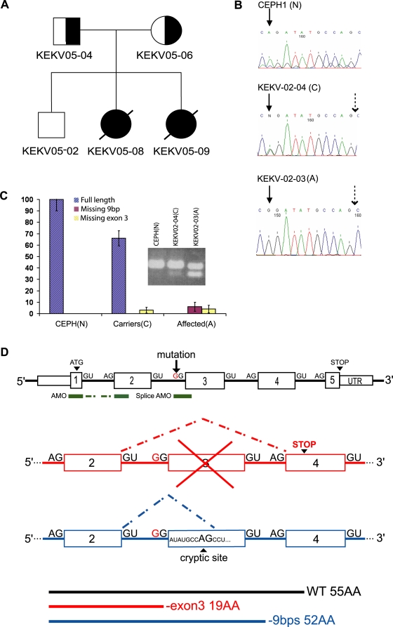
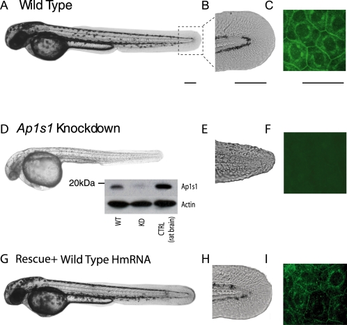
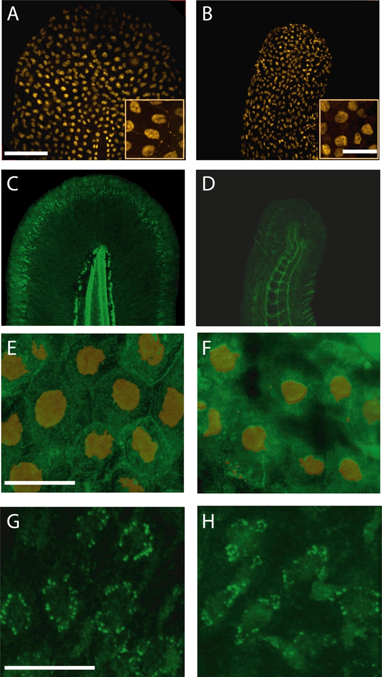
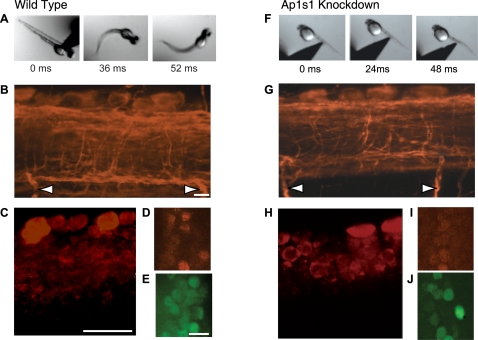
Adaptor protein (AP) complexes regulate clathrin-coated vesicle assembly, protein cargo sorting, and vesicular trafficking between organelles in eukaryotic cells. Because disruption of the various subunits of the AP complexes is embryonic lethal in the majority of cases, characterization of their function in vivo is still lacking. Here, we describe the first mutation in the human AP1S1 gene, encoding the small subunit sigma1A of the AP-1 complex. This founder splice mutation, which leads to a premature stop codon, was found in four families with a unique syndrome characterized by mental retardation, enteropathy, deafness, peripheral neuropathy, ichthyosis, and keratodermia (MEDNIK). To validate the pathogenic effect of the mutation, we knocked down Ap1s1 expression in zebrafish using selective antisens morpholino oligonucleotides (AMO). The knockdown phenotype consisted of perturbation in skin formation, reduced pigmentation, and severe motility deficits due to impaired neural network development. Both neural and skin defects were rescued by co-injection of AMO with wild-type (WT) human AP1S1 mRNA, but not by co-injecting the truncated form of AP1S1, consistent with a loss-of-function effect of this mutation. Together, these results confirm AP1S1 as the gene responsible for MEDNIK syndrome and demonstrate a critical role of AP1S1 in development of the skin and spinal cord.


Conflict of interest statement

The authors have declared that no competing interests exist.

Figures

Figure 1
Figure 1. Identification and characterization of a splice mutation in AP1S1.
A) Pedigree of the fourth MEDNIK family from the Kamouraska region. B) Sequence chromatograms of the intron 2/exon 3 junction of AP1S1 in a normal control (N), a carrier (C) and an affected individual (A) from family EKV3-02. The filled arrow indicates the mutation (A to G) and the dotted arrow points the cryptic splice site. C) Expression of the AP1S1 isoforms. Relative expression levels of each mRNA species (scaled to 100% of control values on the y-axis) are shown for normal controls (N; n = 2) carriers (C; n = 6) and affected individuals (A; n = 3). Values were averaged from three independent experiments. 18S RNA was used to normalize the mRNA quantity. The expression levels from this latter mRNA species could not be distinguished from the wild-type in the carriers in this figure. The expression levels are indicated as percentage of the control values Inset: RT-PCR showing the different species observed. The upper band (166 bp) contains the full-length species in both the control (N) and the carrier (C), whereas the lower band (57 bp) corresponds to the species lacking exon 3. A third mRNA species lacking 9bp, generated by the use of a cryptic splice site, was confirmed by sequencing the upper band (157 bp) of the affected individual (A). D) Schematic representation of the Human AP1S1 gene. The A>G mutation in the acceptor splice site of exon 3 predicts skipping of this exon, leading to a premature stop codon. The use of an alternative acceptor splice site within exon 3 results in a mRNA lacking 9 bps coding for an in frame protein. The location of the two different morpholinos used to knockdown Ap1s1 in zebrafish, targeting either ATG or exon 3 acceptor splice site, are shown in green.
Figure 2
Figure 2. Morphological phenotype of Ap1s1 knockdown zebrafish is rescued by over expression of human AP1S1.
Transmitted light images of 48 hpf Ap1s1 KD larvae show their smaller size, reduced pigmentation (D) and skin disorganization (E) compared to the WT (A, B) and the rescued larvae (G,H). Immunofluorescence of wholemount zebrafish using anti-Ap1s1 antibody showing localization of Ap1s1 to the plasma membrane (C, polygonal) and to a well defined perinuclear ring, in both normal (C) and rescued larvae (G), whereas only a residual and diffuse staining could be observed in KD larvae (F). Western blot analysis (D, inset) indicates nearly complete knockdown of Ap1s1 protein (WT = wild-type, KD = knockdown, CTRL = control rat brain proteins). To normalize the western blot analysis, proteins extracted from WT, KD and CTRL larvae were incubated with anti-actin. Scale bars in (A, D, G) = 100 µm, (B, C, E, F, H, I-Ciii) = 50 µm.
Figure 3
Figure 3. Ap1s1 knockdown is associated with abnormal distribution of laminin and cadherin.
Despite changes in the size and shape of the tail, p63-labelled nuclei (orange) of basal keratinocytes were present in KD larvae. (B). The insets show enlarged views of small groups of keratinocytes. Immunolabelling for laminin (green) in WT (C) showed normal distribution of the basement membrane along the fin fold margin, whereas in the KD larvae the residual laminin labeling was diffuse and disorganized (D). Compared to WT keratinocytes (E) labeled with p63 (orange), cadherin (green) is decreased at the cell membrane of the KD larvae (F). However, the localization of cytokeratin in WT (G) and in KD larvae (H) appeared similar. Scale bars: 10 µm, Insets in A, B, = 20 µm.
Figure 4
Figure 4. Abnormal behavioral phenotype and impaired development of spinal neural network of Ap1s1 knockdown zebrafish.
Consecutive images from films illustrating the response to touch in 48 hpf wild type (WT) (A) and Ap1s1 KD larvae (F). The WT larva reacted to touch by swimming away. In contrast, the Ap1s1 KD larvae exhibit slow and impaired reaction. Immunostaining in wholemount 48 hpf WT (B–E) and Ap1s1 KD larvae (G–J) illustrating the reduced axonal labeling (anti-acetylated tubulin; B and G red, the arrows point to the ventral roots), the reduced number of newly born cells (anti-HU antibody; red in C and H), and the number of interneurons (anti-Pax2 antibody; E and J green) in the Ap1s1 KD larvae. In turn, the number of motoneurons (anti-HB9 antibody; D and I, red) was similar both in WT and Ap1s1 KD. Scale bars: 25 µM.

References

    1. Bonifacino JS, Glick BS. The mechanisms of vesicle budding and fusion. Cell. 2004;116:153–166. - PubMed
    1. Robinson MS. Adaptable adaptors for coated vesicles. Trends Cell Biol. 2004;14:167–174. - PubMed
    1. Owen DJ, Collins BM, Evans PR. Adaptors for clathrin coats: structure and function. Annu Rev Cell Dev Biol. 2004;20:153–191. - PubMed
    1. Boehm M, Bonifacino JS. Adaptins: the final recount. Mol Biol Cell. 2001;12:2907–2920. - PMC - PubMed
    1. Deborde S, Perret E, Gravotta D, Deora A, Salvarezza S, et al. Clathrin is a key regulator of basolateral polarity. Nature. 2008;45:2719–723. - PMC - PubMed

Publication types

MeSH terms

Substances