

Discriminating neurogenic from myopathic disease via measurement of muscle anisotropy
- PMID: 19058193
- PMCID: PMC2719295
- DOI: 10.1002/mus.21115
Discriminating neurogenic from myopathic disease via measurement of muscle anisotropy
Abstract
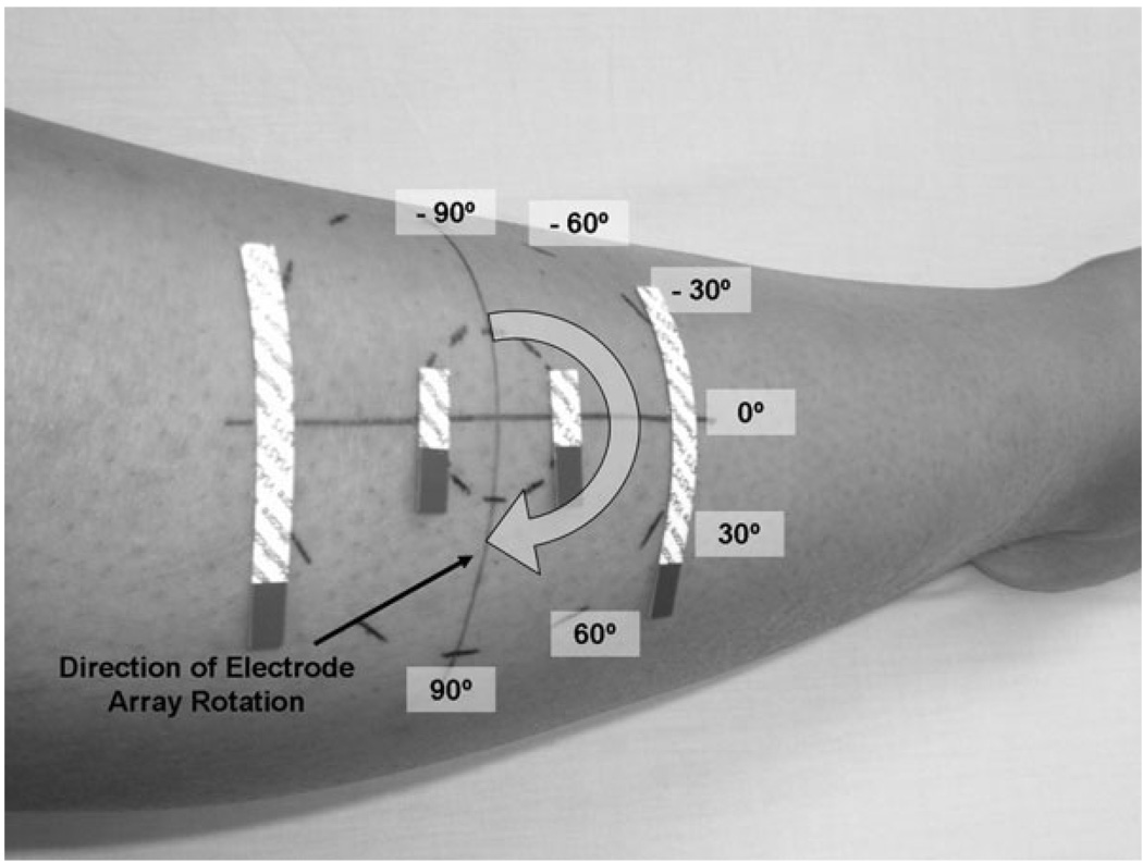
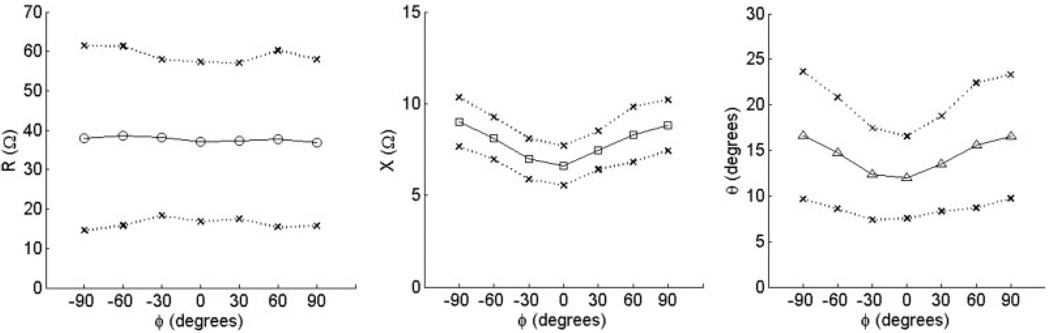
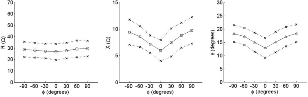
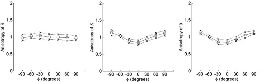
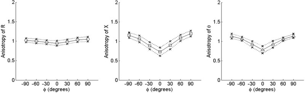
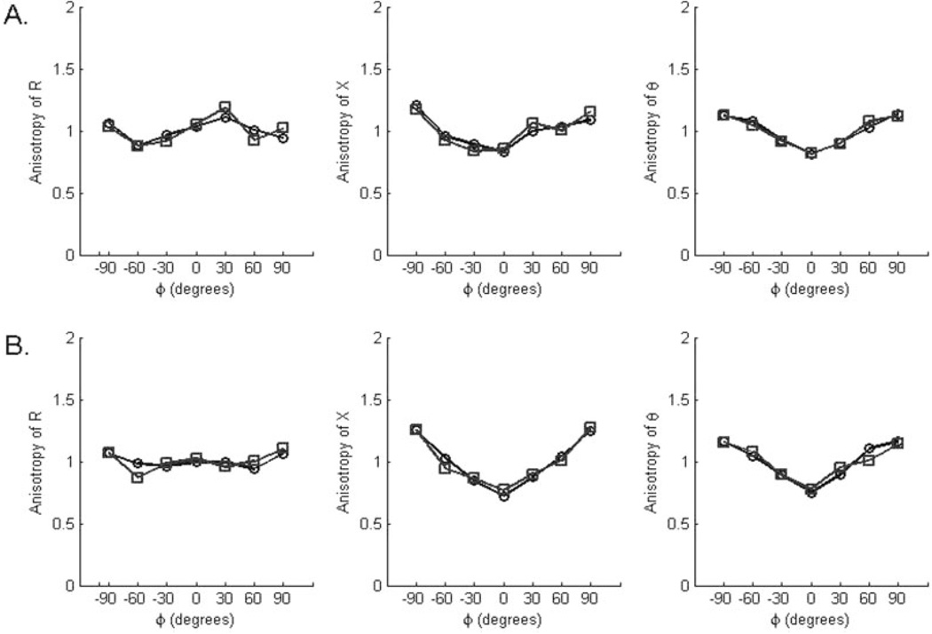
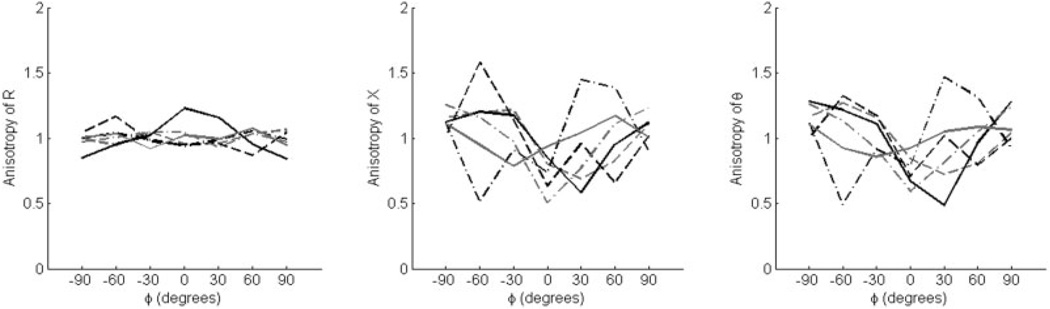
Skeletal muscle is electrically anisotropic, with a tendency for applied electrical current to flow more readily along muscle fibers than across them. In this study, we assessed a method for non-invasive measurement of anisotropy to determine its potential to serve as a new technique for distinguishing neurogenic from myopathic disease. Measurements were made on the biceps brachii and tibialis anterior muscles in 15 normal subjects and 12 patients with neuromuscular disease (6 with amyotrophic lateral sclerosis and 6 with various myopathies) using 50 kHZ applied current. Consistent multi-angle anisotropic patterns were found for reactance and phase in both muscles in normal subjects. Normalized anisotropy differences for each subject were defined, and group average values identified. The amyotrophic lateral sclerosis (ALS) patients demonstrated increased and distorted anisotropy patterns, whereas myopathic patients demonstrated normal or reduced anisotropy. These results suggest that non-invasive measurement of muscle anisotropy has potential for diagnosis of neuromuscular diseases.
(c) 2008 Wiley Periodicals, Inc.
Figures

References
-
- Aaron R, Huang M, Shiffman CA. Anisotropy of human muscle via non-invasive impedance measurements. Phys Med Biol. 1997;42:1245–1262. - PubMed
-
- Burger HC, van Dongen R. Specific electrical resistance of body tissues. Phys Med Biol. 1961;5:431–447. - PubMed
-
- Epstein BR, Foster KR. Anisotropy in the dielectric properties of skeletal muscle. Med Biol Eng Comput. 1983;21:51–55. - PubMed
-
- Esper GJ, Shiffman CA, Aaron R, Lee KS, Rutkove SB. Assessing neuromuscular disease with multifrequency electrical impedance myography. Muscle Nerve. 2006;34:595–602. - PubMed
Publication types
MeSH terms
Grants and funding
LinkOut - more resources
Full Text Sources
Other Literature Sources
Medical
Miscellaneous
