. 2009 Jul;48(7):599-609.
doi: 10.1002/mc.20498.

The human promyelocytic leukemia protein is a tumor suppressor for murine skin carcinogenesis

Affiliations

The human promyelocytic leukemia protein is a tumor suppressor for murine skin carcinogenesis

Victoria M Virador et al. Mol Carcinog. 2009 Jul.

Abstract

Expression of the PMLRARalpha fusion dominant-negative oncogene in the epidermis of transgenic mice resulted in spontaneous skin tumors attributed to changes in both the PML and RAR pathways [Hansen et al., Cancer Res 2003; 63:5257-5265]. To determine the contribution of PML to skin tumor susceptibility, transgenic mice were generated on an FVB/N background, that overexpressed the human PML protein in epidermis and hair follicles under the control of the bovine keratin 5 promoter. PML was highly expressed in the epidermis and hair follicles of these mice and was also increased in cultured keratinocytes where it was confined to nuclear bodies. While an overt skin phenotype was not detected in young transgenic mice, expression of keratin 10 (K10) was increased in epidermis and hair follicles and cultured keratinocytes. As mice aged, they exhibited extensive alopecia that was accentuated on the C57BL/6J background. Following skin tumor induction with 7, 12-dimethylbenz[a]anthracene (DMBA) as initiator and 12-O-tetradecanoylphorbol-13-acetate (TPA) as promoter, papilloma multiplicity and size were decreased in the transgenic mice by 35%, and the conversion of papillomas to carcinomas was delayed. Cultured transgenic keratinocytes underwent premature senescence and upregulated transcripts for p16 and Rb but not p19 and p53. Together, these changes suggest that PML participates in regulating the growth and differentiation of keratinocytes that likely influence its activity as a suppressor for tumor development.


Figures
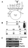
Fig. 1
Fig. 1
A. Human PML (PML-1 M73778) was cloned downstream of the K5 regulatory region, into the 3’ end of the β-globin intron in a construct containing the bovine keratin 5 promoter, the rabbit β-globin intron and an SV40 Poly(A) termination signal [19]. B. Southern blot analysis of DNA from founder lines I1, J1, H4, H1, and G6. Wt represents a full length PML probe prepared by digestion of pcDNA/PML with Not1. C. Genotyping by PCR of a typical litter of K5-PMLH4, testing consistently positive for all primer sets.
Fig. 2
Fig. 2
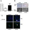
A. Immunohistochemistry of paraffin embedded skin sections of mice from line K5-PMLG6 and control FVB/N stained for PML. B. Immunofluorescence of keratinocytes cultured 4 days in medium containing 0.05 mM Ca 2+, from line K5-PML H4 and control FVB/N with anti-PML (H238, Santa Cruz). Localization is observed in nucleus (arrow) identified by DAPI staining. C. Immunoblots in primary cultured keratinocytes from the H4 line and control littermates cultured for 4 days in medium containing increasing calcium concentration (0.05–0.2 mM).
Fig. 3
Fig. 3
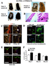
K5-PML mice develop permanent alopecia. A. Eleven and sixteen month old male and five month old female K5-PML mice and littermates are shown. Mice were F2 crosses K5-PML (FVB/N) × C57BL/6J. Figures are labeled tg and wt for simplicity. B. Hematoxylin-eosin staining of a margin between haired and alopecic skin (40x). Right panel shows a keratinized follicle (arrow) in an alopecic area (200x). C. K5-PML H4 transgenic mice express the differentiation marker K10 more extensively than wild type littermates. Ethanol fixed skin sections of wild type and transgenic littermates as newborns and 20 days pp were stained with antibodies to K14 (green) to mark the basal cells, and K10 (red) to visualize differentiated cells. Arrow denotes K10 staining in anagen hair follicles in K5-PML skin. D. Primary keratinocytes grown in medium containing 0.05 mM or 0.2 mM calcium were immunostained to detect K10 expression and nuclei were labeled with DAPI. E. Skin sections from two K5-PML and FVB/N littermates were immunostained for K10 showing areas of alopecia with extension of K10 expression into the hair follicle in the transgenic skin. F. Percent PCNA positive epidermal cells in skin during the first hair cycle. Basal interfollicular cells and those in the upper portion of the hair canal were counted excluding the dermal papilla area. Five random fields were scored for each section.
Fig. 4
Fig. 4
Induction of skin tumors by chemical carcinogens. A. Fifteen mice per group were treated once with DMBA, followed by weekly applications of TPA. A. (left) papilloma incidence (percent tumor-bearing mice) counted between weeks 10 and 22 post-DMBA treatment, and (right) papilloma multiplicity (number of papillomas per mouse), was calculated for each time point. By two way ANOVA, the difference between both curves is significant (p<0.0001) in the case of papilloma multiplicity but not in the case of papilloma incidence. B. Papilloma and carcinoma sizes (mm3) were measured in remaining tumors upon termination of the experiment. Scatter plots represent all tumor sizes and their mean. T test of the mean values was not significant for papillomas and p= 0.04 for carcinoma sizes. C. Conversion to carcinoma was delayed approximately 5 weeks in K5-PML transgenic mice. Log-rank test of the differences between groups was significant at p=0.04.
Fig. 5
Fig. 5
A. Keratinocytes from K5-PML newborn mice senesce after 9 days in culture. Total and SA-β-galactosidase positive cells [24] were scored. Two random fields were scored per well in a 24 well plate and the experiment was repeated twice. *** indicates p=0.01. B. Top, representative images of SA-β-gal staining of wild type and transgenic keratinocytes of animals of the FVB/N background at 10 days in culture. Bottom panels show phase contrast morphology of keratinocytes from K5-PML or wildtype littermates of the C57BL6/J background cultured for 9 days. C. Cultures of wildtype and transgenic keratinocytes were immunostained for p16 (green) and DAPI (blue) at day 1 and day 9 in culture.
Fig. 6
Fig. 6
Quantitative real time PCR analysis of keratinocyte transcripts. K5-PML and FVB/N keratinocytes were isolated and cultured for indicated times prior to RNA isolation. Quantitative PCR was performed as described in Materials and Methods and presented relative to the GAPDH control. The experiments were repeated two times on independent cultures isolated from different mouse litters.

References

    1. Hansen LA, Brown D, Virador V, et al. A PMLRARA transgene results in a retinoid-deficient phenotype associated with enhanced susceptibility to skin tumorigenesis. Cancer Res. 2003;63(17):5257–5265. - PubMed
    1. Melnick A, Licht JD. Deconstructing a disease: RARalpha, its fusion partners, and their roles in the pathogenesis of acute promyelocytic leukemia. Blood. 1999;93(10):3167–3215. - PubMed
    1. Everett RD. DNA viruses and viral proteins that interact with PML nuclear bodies. Oncogene. 2001;20(49):7266–7273. - PubMed
    1. Koken MH, Linares-Cruz G, Quignon F, et al. The PML growth-suppressor has an altered expression in human oncogenesis. Oncogene. 1995;10(7):1315–1324. - PubMed
    1. Gurrieri C, Capodieci P, Bernardi R, et al. Loss of the tumor suppressor PML in human cancers of multiple histologic origins. Journal of the National Cancer Institute. 2004;96(4):269–279. - PubMed

Publication types

MeSH terms