Review
. 2008 Dec;18(6):658-66.
doi: 10.1016/j.sbi.2008.11.005.

Cobalamin-dependent and cobamide-dependent methyltransferases

Affiliations
Review

Cobalamin-dependent and cobamide-dependent methyltransferases

Rowena G Matthews et al. Curr Opin Struct Biol. 2008 Dec.

Abstract

Methyltransferases that employ cobalamin cofactors, or their analogs the cobamides, as intermediates in catalysis of methyl transfer play vital roles in energy generation in anaerobic unicellular organisms. In a broader range of organisms they are involved in the conversion of homocysteine to methionine. Although the individual methyl transfer reactions catalyzed are simple S(N)2 displacements, the required change in coordination at the cobalt of the cobalamin or cobamide cofactors and the lability of the reduced Co(+1) intermediates introduces the necessity for complex conformational changes during the catalytic cycle. Recent spectroscopic and structural studies on several of these methyltransferases have helped to reveal the strategies by which these conformational changes are facilitated and controlled.


Figures

Figure 1
Figure 1. (A) The structure of methylcobalamin
The cobalt in methylcobalamin, which is formally in the +3 oxidation state, is coordinated by four equatorial ligands contributed by the corrin ring and by two axial ligands. The β-axial ligand (the upper axial ligand in the orientation shown here) is the methyl anion in methylcobalalamin, while the lower axial ligand (the α ligand) in the free cofactor is a dimethylbenzimidazole nucleotide substituent of the D-ring of the corrin [32]. (B) The structure of methylcobalamin in the cobalamin-binding module of methionine synthase [6]. The dimethyl-benzimidazole nucleotide has dissociated, and cobalt is coordinated by His759 from the protein. His 759 is linked by a network of hydrogen bonds to Asp757 and Ser810. A signature Asp-X-His-X-X-Gly----Ser-Leu motif is diagnostic of the His-on state of cobalamin in many corrinoid proteins. While methylcobalamin is preferentially six-coordinate, cob(I)alamin is planar four-coordinate, due to the electron density of the lone pair of electrons that remain after transfer of the methyl group as a carbocation. As the cofactor cycles in catalysis between methylcobalamin and cob(I)alamin forms, the changes in preferred coordination geometry require that histidine dissociation be coupled with methyl transfer [33]. This histidine dissociation is accompanied by uptake of a proton, presumably due to protonation of the His-Asp-Ser ligand triad [34]. (C) The structure of cobalamin in the reactivation conformation of methionine synthase [10••]. In this structure Nε of His759 has rotated away from the cobalt of the cofactor, and is now 5.6 Å displaced from its position in (B). The structures in B and C are similarly oriented with respect to the cobalamin-binding domain while those in A and C are aligned based on the corrin rings. D. Cartoon of the change in cobalt coordination required for reduction of cob(II)alamin to cob(I)alamin and subsequent alkylation in His-on methyltransferases. Reactivation involves reduction of cob(II)alamin to cob(I)alamin and methylation of cob(I)alamin to regenerate methylcobalamin. In methionine synthase flavodoxin serves as the electron donor [35], and adenosylmethionine serves as the methyl donor for reactivation [7]. Cob(II)alamin is preferentially five-coordinate, and in methionine synthase His759 is coordinated in the α-axial position. Its reduction to cob(I)alamin is facilitated by dissociation of the histidine ligand. Binding of flavodoxin to the cob(II)alamin form of the enzyme results in dissociation of His759 from the cobalt, and uptake of a proton [36]. Following reduction to cob(I)alamin, methyl transfer from adenosylmethionine initially results in the formation of His-off methylcobalamin and the rate-limiting step in reactivation is the appearance of His-on methylcobalamin [37].
Figure 2
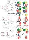
Figure 2. The catalytic cycles and proposed conformations of three well-studied methyltransferases
(A) Methionine synthase The cobalamin cofactor is alternately methylated by CH3-H4folate bound to the Fol domain (b, requiring the Fol:Cob conformation cartooned on the right) and demethylated by homocysteine bound to the Hcy domain (a, Hcy:Cob). Occasionally, the cob(I)alamin cofactor undergoes oxidation. Return of the inactive cob(II)alamin species to the catalytic cycle require a reductive reactivation, in which adenosylmethionine bound to the C-terminal domain (c, AdoMet:Cob) serves as the methyl donor [7]. In E. coli, flavodoxin serves as the source of electrons for reductive activation [35], while in humans the source of electrons is methionine synthase reductase, a protein with homology to both flavodoxin and NADPH-ferredoxin (flavodoxin) oxidoreductase [38,39]. The remaining conformation (d, Cap:Cob) has only been seen in crystals of the isolated cobalamin-binding module. While the cartoon shows all four domains, x-ray structures of the full length protein have never been obtained, and the cartoons are based on docking of the N-terminal and C-terminal halves of the protein in a plausible manner. (B) Acetyl-CoA synthase. The 5-hydroxymethylbenzimidazolyl cobamide cofactor of the corrinoid iron/sulfur protein (AcsCD) is alternately methylated by CH3-H4folate bound to the AcsE methyltransferase (b, Fol:Cob) and demethylated by transfer of the methyl group to Ni+1 in acetyl CoA synthase (a, ACS:Cob). When the cob(I)amide undergoes occasional oxidation to cob(II)amide, it is returned to the catalytic cycle by reduction by the Fe4-S4 cluster on the large subunit (AcsC) of the corrinoid iron/sulfur protein (c, Fe-S:Cob). This cluster is in turn re-reduced with electrons derived from ferredoxin or from enzymes coupled to ferredoxin. The remaining conformation (d, Cap:Cob) is the one seen in the isolated corrinoid iron-sulfur protein [18••]). (C) Methanol:coenzyme M methyltransferase. The corrinoid-binding protein MtaC forms a complex with MtaA (the coenzyme M-binding methyltransferase) and MtaB (the methanol-binding methyltransferase). During catalysis the complex cycles between CoM:Cob (a) and Methanol:Cob (b) conformations. The latter complex has been characterized crystallo-graphically in the absence of MtaA. Activation of the cob(II)amide cofactor requires reduction by ferredoxin and is coupled to ATP hydrolysis by the action of the MAP protein (c, ATP:Cob). The Cap:Cob conformation (d) is hypothetical.
Figure 3
Figure 3. (A) Structure of the reactivation (AdoMet:Cob) conformation of methionine synthase[10••]
This structure was obtained with a fragment of the enzyme containing only the cobalamin-binding and adenosylmethionine-binding modules. The AdoMet-binding module is shown in blue, the cobalamin-binding domain in red, and the cap in tan. To reduce the conformational flexibility yet at the same time keeping the active site intact, a strategy of disulfide cross-linking between the two modules was used. Two cysteine mutations, Ile690Cys (in the cap domain) and Gly743Cys (in the cobalamin-binding domain) were introduced. The resulting disulfide cross-link sufficiently favored the conversion from His-on to His-off such that the protein crystallized in the AdoMet:Cob conformation. Nε of His-759 has moved 5.6 Å away from the cobalt towards the AdoMet binding module and is involved in an intermodular hydrogen bonding contact. (B) Structure of the MtaBC complex [27••] of methanol:coenzyme M methyltranferase, shown in the same orientation as methionine synthase in (A). MtaB forms a decorated TIM barrel (yellow), with the zinc atom (gray sphere) located at the C-terminal opening of the barrel in close proximity to the corrinoid of MtaC. The zinc is ligated by two cysteines and a glutamate, while the identity of the fourth ligand remains unclear. The sequences containing these three ligands bear no resemblance to sequences associated with zinc binding in other family members, despite the fact that the closest structural relative of MtaB is the homocysteine-binding domain of methionine synthase. Methanol is not present in the MtaBC complex, but if coordinated to the zinc by its hydroxyl oxygen, could be positioned appropriately for methyl transfer to the cobalt of the 5-hydroxybenzimidazolyl cobamide of MtaC, MtaC consists of two domains, a corrinoid-binding domain (red) and a four helix bundle or cap (tan) with an N-terminal extension (light blue). (C) Details of the intermodular interaction of His-off methionine synthase. His759 forms a hydrogen bond to Glu1069 and a water mediated hydrogen bond with Asp1093. This contact is expected to stabilize the His-off form in the AdoMet:Cob conformation.
Figure 4
Figure 4. (A) The homocysteine binding domain of methionine synthase in the presence or absence of homocysteine[14**]
This panel shows a tructure superposition of the MetH resting state (gray) and the Hcy bound state (green) with Zn(R) representing the zinc atom at the resting state (no substrates bound) and Zn(H) the zinc atom at the Hcy bound state. Binding of homocysteine leads to an inversion of geometry at the active-site zinc and the displacement of Asn234 from the zinc coordination sphere. The zinc moves 2 Å on binding of homocysteine. (B) The CH3-H4folate-binding domain of the corrinoid iron-sulfur protein methyltransferase AcsE [41•]. CH3-H4folate is unprotonated in this structure and N5 accepts a hydrogen bond from Asn199.

Similar articles

Cited by

References

    1. Matthews RG. Corrinoid- and cobalamin-dependent methyltransferases. In: Sigal A, Sigal H, Sigal RKO, editors. Metal-carbon bonds in enzymes and cofactors. Royal Society of Chemistry; 2009:in press. vol 6 of “Metal Ions in Life Sciences”.
    1. Croft MT, Lawrence AD, Raux-Deery E, Warren MJ, Smith AG. Algae acquire vitamin B12 through a symbiotic relationship with bacteria. Nature. 2005;438:90–93. - PubMed
    1. Galagan JE, Nusbaum C, Roy A, Endrizzi MG, Macdonald P, FitzHugh W, Calvo S, Engels R, Smirnov S, Atnoor D, et al. The genome of M. acetivorans reveals extensive metabolic and physiological diversity. Genome Res. 2002;12:532–542. - PMC - PubMed
    1. Goulding CW, Postigo D, Matthews RG. Cobalamin-dependent methionine synthase is a modular protein with distinct regions for binding homocysteine, methyltetrahydrofolate, cobalamin, and adenosylmethionine. Biochemistry. 1997;36:8082–8091. - PubMed
    1. Evans JC, Huddler DP, Hilgers MT, Romanchuk G, Matthews RG, Ludwig ML. Structures of the N-terminal modules imply large domain motions during catalysis by methionine synthase. Proc Natl Acad Sci USA. 2004;101:3729–3736. - PMC - PubMed

Publication types

MeSH terms

LinkOut - more resources