Iron homeostasis and eye disease
- PMID: 19059309
- PMCID: PMC2718721
- DOI: 10.1016/j.bbagen.2008.11.001
Iron homeostasis and eye disease
Abstract
Background: Iron is necessary for life, but excess iron can be toxic to tissues. Iron is thought to damage tissues primarily by generating oxygen free radicals through the Fenton reaction.
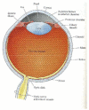
Methods: We present an overview of the evidence supporting iron's potential contribution to a broad range of eye disease using an anatomical approach.
Results: Iron can be visualized in the cornea as iron lines in the normal aging cornea as well as in diseases like keratoconus and pterygium. In the lens, we present the evidence for the role of oxidative damage in cataractogenesis. Also, we review the evidence that iron may play a role in the pathogenesis of the retinal disease age-related macular degeneration. Although currently there is no direct link between excess iron and development of optic neuropathies, ferrous iron's ability to form highly reactive oxygen species may play a role in optic nerve pathology. Lastly, we discuss recent advances in prevention and therapeutics for eye disease with antioxidants and iron chelators.
General significance: Iron homeostasis is important for ocular health.
Figures


Similar articles
-
Oxidative stress and iron homeostasis: mechanistic and health aspects.Crit Rev Clin Lab Sci. 2008;45(1):1-23. doi: 10.1080/10408360701713104. Crit Rev Clin Lab Sci. 2008. PMID: 18293179 Review.
-
The role of antioxidants and iron chelators in the treatment of oxidative stress in thalassemia.Ann N Y Acad Sci. 2010 Aug;1202:10-6. doi: 10.1111/j.1749-6632.2010.05577.x. Ann N Y Acad Sci. 2010. PMID: 20712766
-
Iron metabolism and toxicity.Toxicol Appl Pharmacol. 2005 Jan 15;202(2):199-211. doi: 10.1016/j.taap.2004.06.021. Toxicol Appl Pharmacol. 2005. PMID: 15629195 Review.
-
Iron homeostasis and toxicity in retinal degeneration.Prog Retin Eye Res. 2007 Nov;26(6):649-73. doi: 10.1016/j.preteyeres.2007.07.004. Epub 2007 Aug 11. Prog Retin Eye Res. 2007. PMID: 17921041 Free PMC article. Review.
-
The Role of the Reactive Oxygen Species and Oxidative Stress in the Pathomechanism of the Age-Related Ocular Diseases and Other Pathologies of the Anterior and Posterior Eye Segments in Adults.Oxid Med Cell Longev. 2016;2016:3164734. doi: 10.1155/2016/3164734. Epub 2016 Jan 10. Oxid Med Cell Longev. 2016. PMID: 26881021 Free PMC article. Review.
Cited by
-
Extraction of Iron from the Rabbit Anterior Chamber with Reverse Iontophoresis.J Ophthalmol. 2015;2015:425438. doi: 10.1155/2015/425438. Epub 2015 Jul 15. J Ophthalmol. 2015. PMID: 26257921 Free PMC article.
-
Gene transcription profile of the detached retina (An AOS Thesis).Trans Am Ophthalmol Soc. 2009 Dec;107:343-82. Trans Am Ophthalmol Soc. 2009. PMID: 20126507 Free PMC article.
-
MicroRNA Expression Analysis in Serum of Patients with Congenital Hemochromatosis and Age-Related Macular Degeneration (AMD).Med Sci Monit. 2017 Aug 22;23:4050-4060. doi: 10.12659/msm.902366. Med Sci Monit. 2017. PMID: 28827515 Free PMC article.
-
The oral iron chelator deferiprone protects against iron overload-induced retinal degeneration.Invest Ophthalmol Vis Sci. 2011 Feb 16;52(2):959-68. doi: 10.1167/iovs.10-6207. Invest Ophthalmol Vis Sci. 2011. PMID: 21051716 Free PMC article.
-
A Novel Ferroptosis Inhibitor UAMC-3203, a Potential Treatment for Corneal Epithelial Wound.Pharmaceutics. 2022 Dec 29;15(1):118. doi: 10.3390/pharmaceutics15010118. Pharmaceutics. 2022. PMID: 36678747 Free PMC article.
References
-
- Anderson G. Mechanisms of iron loading and toxicity. Am J of Hepatology. 2007;82(S12):1128–1131. - PubMed
-
- Wong R, et al. Iron toxicity as a potential factor in AMD. Retina. 2007;27:997–1003. - PubMed
-
- Fleming MD, et al. Microcytic anaemia mice have a mutation in Nramp2, a candidate iron transporter gene. Nat Genet. 1997;16(4):383–6. - PubMed
-
- Abboud S, Haile DJ. A novel mammalian iron-regulated protein involved in intracellular iron metabolism. J Biol Chem. 2000;275(26):19906–12. - PubMed
Publication types
MeSH terms
Substances
Grants and funding
LinkOut - more resources
Full Text Sources
Other Literature Sources
Medical

