. 2009 Feb;1789(2):88-98.
doi: 10.1016/j.bbagrm.2008.11.002. Epub 2008 Nov 14.

TGF-beta1 modulates focal adhesion kinase expression in rat intestinal epithelial IEC-6 cells via stimulatory and inhibitory Smad binding elements

Affiliations

TGF-beta1 modulates focal adhesion kinase expression in rat intestinal epithelial IEC-6 cells via stimulatory and inhibitory Smad binding elements

Mary F Walsh et al. Biochim Biophys Acta. 2009 Feb.

Abstract

TGF-beta and FAK modulate cell migration, differentiation, proliferation and apoptosis, and TGF-beta promotes FAK transcription in intestinal epithelial cells via Smad-dependent and independent pathways. We utilized a 1320 bp FAK promoter-luciferase construct to characterize basal and TGF-beta-mediated FAK gene transcription in IEC-6 cells. Inhibiting JNK or Akt negated TGF-beta-stimulated promoter activity; ERK inhibition did not block the TGF-beta effect but increased basal activity. Co-transfection with Co-Smad4 enhanced the TGF-beta response while the inhibitory Smad7 abolished it. Serial deletions sequentially removing the four Smad binding elements (SBE) in the 5' untranslated region of the promoter revealed that the two most distal SBE's are positive regulators while SBE3 exerts a negative influence. Mutational deletion of two upstream p53 sites enhanced basal but did not affect TGF-beta-stimulated increases in promoter activity. TGF-beta increased DNA binding of Smad4, phospho-Smad2/3 and Runx1/AML1a to the most distal 435 bp containing 3 SBE and 2 AML1a sites by ChIP assay. However, although point mutation of SBE1 ablated the TGF-beta-mediated rise in SV40-promoter activity, mutation of AML1a sites did not. TGF-beta regulation of FAK transcription reflects a complex interplay between positive and negative non-Smad signals and SBE's, the last independent of p53 or AML1a.


Figures

Figure 1
Figure 1
TGF-β enhances FAK protein expression and FAK promoter activity. (A) Subconfluent IEC-6 cells were treated with 1ng/ml TGF-β1 for 24 hours. Cell lysates were analyzed by Western blot for total FAK (tFAK) and tubulin as loading control. Data, normalized to tubulin, is presented as percent of control non-treated cells. Densitometric analysis (Ave ± SEM) of 4 separate experiments is depicted in the bars; a representative Western blot is shown above. Asterisk denotes significant difference (p<0.05) from control, untreated cells. (B) Agarose gel of the 1.3 kb construct containing the 5’ flanking region of the human FAK promoter. The fragment was subcloned into a pGL2 Basic vector to generate a FAK-promoter-luciferase construct. (C) Cells transfected with the control empty vector (pGL2) or the FAK promoter construct (wt.FAK) were exposed to 1ng/ml TGF-β1 for 24 hours prior to analysis via a Dual Luciferase® Reporter Assay System. Promoter luciferase activity is express as the ratio of luciferase to that of a co-transfected pRL-TK control vector encoding Renilla luciferase. Asterisk denotes significant fold increase over pGL2 empty vector, pound sign over untreated wt.FAK construct (p<0.05).
Figure 2
Figure 2
Short-term exposure (1 hr) to TGF-β stimulates phosphorylation/activation of several non-Smad signaling pathways. IEC-6 cells were pretreated for 1 hr with the MEK inhibitor PD98059 (A), the PI-3K (phospho-inositol-3-kinase) inhibitor, LY294002 (B), JNK inhibitor II (JNK Inh II) (C) (each, 10 µM), or an equivalent concentration of the DMSO vehicle prior to 1 hr stimulation with I ng/ml TGF-β (black bars). Cell lysates were analyzed by Western blot with specific antibodies for phospho-ERK (panel A), phospho-Akt (Thr308: panel B) or phospho-JNK (Thr183/Tyr185; panel C) and with antibodies specific to GAPDH as a control. Bar graphs represent densitometric analysis of each specific phosphoprotein signal as a ratio of the GAPDH control; representative Western blots are depicted above the bars. Bars show percent increase over respective control; asterisks indicate statistical significance (p<0.03) from DMSO-treated controls, calculated from the original signal/GAPDH ratios.
Figure 3
Figure 3
ERK signals exert a significant inhibitory effect on basal FAK promoter activity while PI-3K and JNK signal activation mediates TGF-β stimulation of activity. Cells were co-transfected with the wt.FAK promoter-luciferase and Renilla- luciferase constructs 24 hours prior to pre-treatment with the TGF-β receptor antagonist SB431542 (0.5 µM), the MEK inhibitor PD98059 (10 µM), the PI-3K inhibitor LY294002 (10 µM), or JNK Inhibitor II (JNK Inh; 10 µM) for 1 hr. Control cells were treated with an equivalent concentration of the DMSO vehicle (open bar) or the requisite inhibitor (grey bars), except for JNK Inhibitor II where the inactive (negative) control peptide (Neg Cont; 10 µM) was added. Cells were then exposed to 1 ng/ml TGF-β (black bars) for 24 hours before lysis and luciferase assay. Results are presented as percent of the DMSO control (Ave ± SEM of 3–9 experiments); statistical comparisons were calculated from triplicate averages of the original luciferase/renilla ratios. Asterisks denote statistical difference in promoter activity over the DMSO control (p<0.001 for TGF-β treatment alone; p<0.002 for PD98059 alone). Pound signs indicate statistical differences of TGF-β-treated cells over their respective controls (p<0.05 for PD98059 and Negative JNK control).
Figure 4
Figure 4
The activating Co-Smad Smad4 increases FAK promoter activity in response to TGF-β while the inhibitory Smad7 represses it. IEC-6 cells were co-transfected with a vector containing the FAK promoter-luciferase construct (wt.FAK) or FAK-pcDNA3, FLAG- Smad4 and Smad7 PcDNA or both the Smad4 and Smad7 constructs along with the internal renilla control pRL-TK vector. Cells were incubated for 24 hours for expression of the desired protein then treated with 1ng/ml TGF-β for a further 24 hours before lysis and analysis of luciferase activity. Results are presented as fold increase of the luciferase/renilla ratio over the empty vector controls which were unchanged by transfection with any of the constructs (not shown). Asterisks show significant differences between TGF-β-treated cells (black bars) and their respective controls (open bars; p<0.05 for each).
Figure 5
Figure 5
Decreasing Smad2 expression via specific siRNA has no effect on basal FAK promoter activity. IEC-6 cells were transfected with a specific siRNA pool to Smad2 (siSmad2; black bars) or a non-targeting control sequence (NT1; open bars). After 24 hours, the wtFAK-luciferase and renilla/luciferase constructs were introduced into half the cells and all cells incubated a further 24 hours. Corresponding cells were then lysed for either Western blot analysis or luciferase assay. Left panel: Cell lysates were blotted with a specific Smad2 antibody and antibody to GAPDH as control. Bars represent densitometric analysis, expressed as percent of the NT1 control; a representative blot is shown above. A significant difference is denoted by the asterisk (p<0.001 vs NT1). Right panel: Bars depict FAK promoter luciferase activity of the siSmad2-treated cells (black bar) calculated from luciferase/renilla ratios and expressed as percent of NT1 control-treated cells (open bar).
Figure 6
Figure 6
Smad binding elements (SBE) transduce activation and repression, and modulate TGF-β responsiveness of the FAK promoter. (A) Schematic representation of the generation of FAK promoter constructs with consecutive deletions of 4 SBE sites; +47 is the putative transcription start site. Locations of SBE, p53, NF-κB consensus sequences and forward and reverse primer sites (arrows) are indicated. Each of the seven PCR amplified fragments (FAK-A to FAK-G) were ligated into a promoterless luciferase vector (pGL2) 24 hours before treatment with TGF- β. Cells were lysed for luciferase activity after 24 hours of exposure; activity was normalized to that of the renilla internal control. (B) Basal FAK promoter activity in IEC-6 cells transfected with each of the seven deletion mutants (grey bars) is presented as a percent of the activity of the full-length (wt.FAK/FAK-A) construct (open bar). Asterisk denotes a significant increase and pound signs a significant decrease in basal promoter activity over the full-length construct containing all 4 SBE’s and both p53 and NF-κB sites (p<0.05). (C) TGF-β-stimulated FAK promoter activity (black bars) for the full length (FAK-A) and each deletion mutant construct (FAK-B to FAK-G) was calculated as the ratio of luciferase to the internal rinella control and expressed as percent of respective control (open bars). Data presented are Ave±SEM of three individual experiments. Asterisks indicate significant differences from respective controls; p<0.05 for each.
Figure 7
Figure 7
p53 DNA binding sites exert an inhibitory action on baseline FAK promoter activity. (A) Sites of single and tandem p53 deletions in the 5’ flanking region of the FAK promoter are illustrated. The pGL2 FAK promoter plasmid was used as DNA template for mutation by deletion of 8 bases from p53 sites distal (mt.p53.d) or proximal (mt.p53.p) to the start site (+47), or both (mt.p53.d+p). Cells were treated with 1ng/ml TGF-β for 24 hours before lysis and assessment of promoter activity by luciferase. (B) Basal FAK promoter activity, calculated as the ratio of luciferase/rinella activities for each p53 mutant (grey bars), is shown as a percentage of basal activity of cells transfected with the non-mutated (wt.FAK) construct (open bar). Asterisks denote significant differences from wt.FAK (p<0.02). (C) Normalized luciferase results for wt.FAK and each p53 deletion mutant are presented as fold increase over respective untreated control in IEC-6 cells treated with 1 ng/ml TGF-β for 24 hours (black bars). Bars represent Ave±SEM of four experiments. Asterisks indicate statistical difference from untreated control; all statistical comparisons were calculated using the individual, respective controls.
Figure 8
Figure 8
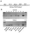
ChIP assay for Smad4, phospho-Smad2/3 and Runx1/AML1 protein binding to the 5’ distal region (−1173 to −738) of the FAK promoter. (A) The primer pair was designed to amplify a 435 bp fragment containing three copies of SBE and two copies of AML1. (B) Chromatin fragments, obtained from cell incubated with and without 1 ng/ml TGF-β for 24 hours, were incubated overnight with A/G Sepharose and antibodies specific for Smad4, phospho-Smad2/3, Runx1 or the IgG control. After elution of the protein-chromatin complexes and phenol:chloroform extraction, the fragments were amplified by PCR using the primer pair. The amplified PCR products for control (top panel) and TGF-β-treated (bottom panel) cells, and control plasmid DNA and antibody lacking (no ab) samples were separated on a 1.2% agarose gel; standard markers are shown in the first and last lane (lanes 1 and 10).
Figure 9
Figure 9
TGF-β responsiveness of the FAK promoter is abolished by point mutation of SBE1. (A) Schematic illustration of point mutations generated in the 130 bp TGF-β responsive region in the 5’ UTR containing one SBE and two AML1a DNA binding sites. The 130 bp (from −1173 to −1043 bases) upstream fragment was PCR amplified and ligated into a pGL2 vector driven by a SV-40 promoter (wt.SBE1). Disabling point mutations were generated in the SBE1 site (pmt.SBE1) or both AML1a sites (pmt.AML1a(d+p)). IEC-6 cells containing each construct were then left untreated (open bars) or treated with 1 ng/ml TGF-β (black bars) for 24 hours. (B) Luciferase activity of the intact SBE1 (wt.SBE1) and point mutated SBE1 (pmt.SBE1) promoters, normalized to the renilla control, is shown as fold increase over the empty pGL2-SV40 vector. (C) Normalized luciferase activity of the pGL2-SV40 vector containing either the intact wt.SBE1 or the mutated AML1a (pmt.AML1a(d+p)) inserts is depicted in the bars as fold increase over the empty vector. Asterisks denote statistical significance compared to respective controls in four separate experiments each (p<0.05 for each).

Similar articles

Cited by

References

    1. Hall PA, Coates PJ, Ansari B, Hopwood D. Regulation of cell number in the mammalian gastrointestinal tract: the importance of apoptosis. J Cell Sci. 1994;107(Pt 12):3569–3577. - PubMed
    1. Sturm A, Dignass AU. Epithelial restitution and wound healing in inflammatory bowel disease. World J Gastroenterol. 2008;14:348–353. - PMC - PubMed
    1. Szabo IL, Pai R, Jones MK, Ehring GR, Kawanaka H, Tarnawski AS. Indomethacin delays gastric restitution: association with the inhibition of focal adhesion kinase and tensin phosphorylation and reduced actin stress fibers. Exp Biol Med (Maywood) 2002;227:412–424. - PubMed
    1. Tarnawski A, Szabo IL, Husain SS, Soreghan B. Regeneration of gastric mucosa during ulcer healing is triggered by growth factors and signal transduction pathways. J Physiol Paris. 2001;95:337–344. - PubMed
    1. Tarnawski AS. Cellular and molecular mechanisms of gastrointestinal ulcer healing. Dig Dis Sci. 2005;50 Suppl 1:S24–S33. - PubMed

Publication types

MeSH terms