

Temporal regulation of Ath5 gene expression during eye development
- PMID: 19059393
- PMCID: PMC2788623
- DOI: 10.1016/j.ydbio.2008.10.046
Temporal regulation of Ath5 gene expression during eye development
Abstract
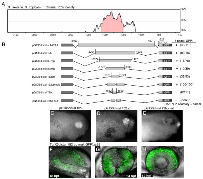
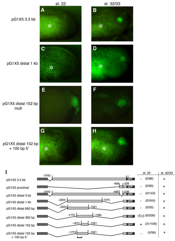
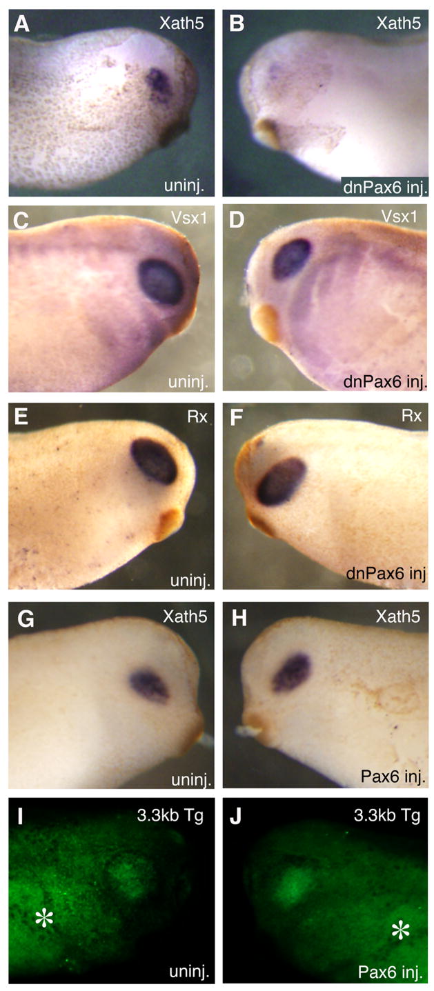
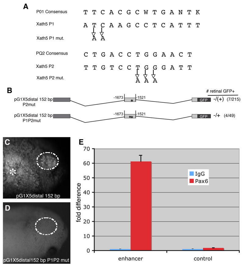
During central nervous system development the timing of progenitor differentiation must be precisely controlled to generate the proper number and complement of neuronal cell types. Proneural basic helix-loop-helix (bHLH) transcription factors play a central role in regulating neurogenesis, and thus the timing of their expression must be regulated to ensure that they act at the appropriate developmental time. In the developing retina, the expression of the bHLH factor Ath5 is controlled by multiple signals in early retinal progenitors, although less is known about how these signals are coordinated to ensure correct spatial and temporal pattern of gene expression. Here we identify a key distal Xath5 enhancer and show that this enhancer regulates the early phase of Xath5 expression, while the proximal enhancer we previously identified acts later. The distal enhancer responds to Pax6, a key patterning factor in the optic vesicle, while FGF signaling regulates Xath5 expression through sequences outside of this region. In addition, we have identified an inhibitory element adjacent to the conserved distal enhancer region that is required to prevent premature initiation of expression in the retina. This temporal regulation of Xath5 gene expression is comparable to proneural gene regulation in Drosophila, whereby separate enhancers regulate different temporal phases of expression.
Figures


Similar articles
-
bHLH-dependent and -independent modes of Ath5 gene regulation during retinal development.Development. 2005 Feb;132(4):829-39. doi: 10.1242/dev.01653. Development. 2005. PMID: 15677728
-
Onset of atonal expression in Drosophila retinal progenitors involves redundant and synergistic contributions of Ey/Pax6 and So binding sites within two distant enhancers.Dev Biol. 2014 Feb 1;386(1):152-64. doi: 10.1016/j.ydbio.2013.11.012. Epub 2013 Nov 16. Dev Biol. 2014. PMID: 24247006 Free PMC article.
-
Math5 encodes a murine basic helix-loop-helix transcription factor expressed during early stages of retinal neurogenesis.Development. 1998 Dec;125(23):4821-33. doi: 10.1242/dev.125.23.4821. Development. 1998. PMID: 9806930
-
Pax6: a multi-level regulator of ocular development.Prog Retin Eye Res. 2012 Sep;31(5):351-76. doi: 10.1016/j.preteyeres.2012.04.002. Epub 2012 May 3. Prog Retin Eye Res. 2012. PMID: 22561546 Review.
-
Eye field specification in Xenopus laevis.Curr Top Dev Biol. 2010;93:29-60. doi: 10.1016/B978-0-12-385044-7.00002-3. Curr Top Dev Biol. 2010. PMID: 20959162 Review.
Cited by
-
All in the family: proneural bHLH genes and neuronal diversity.Development. 2018 May 2;145(9):dev159426. doi: 10.1242/dev.159426. Development. 2018. PMID: 29720483 Free PMC article. Review.
-
Dynamic Polarization of Rab11a Modulates Crb2a Localization and Impacts Signaling to Regulate Retinal Neurogenesis.Front Cell Dev Biol. 2021 Feb 9;8:608112. doi: 10.3389/fcell.2020.608112. eCollection 2020. Front Cell Dev Biol. 2021. PMID: 33634099 Free PMC article.
-
Rbpj direct regulation of Atoh7 transcription in the embryonic mouse retina.Sci Rep. 2018 Jul 5;8(1):10195. doi: 10.1038/s41598-018-28420-y. Sci Rep. 2018. PMID: 29977079 Free PMC article.
-
Distinct effects of Hedgehog signaling on neuronal fate specification and cell cycle progression in the embryonic mouse retina.J Neurosci. 2009 May 27;29(21):6932-44. doi: 10.1523/JNEUROSCI.0289-09.2009. J Neurosci. 2009. PMID: 19474320 Free PMC article.
-
Development of the retina and optic pathway.Vision Res. 2011 Apr 13;51(7):613-32. doi: 10.1016/j.visres.2010.07.010. Epub 2010 Jul 18. Vision Res. 2011. PMID: 20647017 Free PMC article. Review.
References
-
- Amaya E, Musci TJ, Kirschner MW. Expression of a dominant negative mutant of the FGF receptor disrupts mesoderm formation in Xenopus embryos. Cell. 1991;26:257–270. - PubMed
-
- Baker NE, Yu S, Han D. Evolution of proneural atonal expression during distinct regulatory phases in the developing Drosophila eye. Curr Biol. 1996;6:1290–1301. - PubMed
-
- Blader P, Plessy C, Strahle U. Multiple regulatory elements with spatially and temporally distinct activities control neurogenin1 expression in primary neurons of the zebrafish embryo. Mech Dev. 2003;120:211–218. - PubMed
-
- Brown NL, Kanekar S, Vetter ML, Tucker PK, Gemza DL, Glaser T. Math5 encodes a murine basic helix-loop-helix transcription factor expressed during early stages of retinal neurogenesis. Development. 1998;125:4821–4833. - PubMed
Publication types
MeSH terms
Substances
Grants and funding
LinkOut - more resources
Full Text Sources
Medical
Molecular Biology Databases
