

Impact of very old age on the expression of cervical spinal cord cell markers in rats
- PMID: 19059476
- PMCID: PMC2674535
- DOI: 10.1016/j.jchemneu.2008.11.001
Impact of very old age on the expression of cervical spinal cord cell markers in rats
Abstract
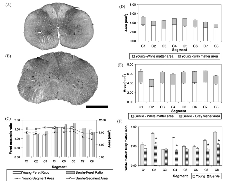
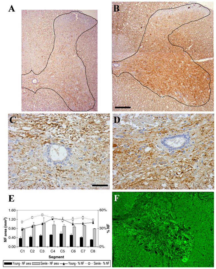
Aging is a process associated with both anatomical changes and loss of expression of some cell markers. Intermediate filaments are known to impart mechanical stability to cells and tissues. Some of them are present in different cell populations of the central nervous system. In order to explore the impact of extreme age we immunohistochemically characterized the changes in intermediate filaments and other cellular markers present in cells populating the gray matter cervical spinal cord of very old rats (28 months) taking young (5 months) counterparts as a reference. The spinal cord weight of the senile animals (12.6+/-1.1 g) was significantly higher (P<0.001) than that of the young animals (8.4+/-1.1 g). Spinal cord length also increased significantly (P<0.05) with age (7.9+/-0.3 cm vs. 8.28+/-0.1 cm for young and senile, respectively). An increase in both neurofilament staining area and density was observed in senile rats in comparison to young animals. A significant (P<0.05) age-related increment in the mean area of the cervical segments was observed. Vimentin expression in the ependymal zone decreased in area and intensity during aging. Our data show that there are some significant changes in the morphological and histochemical patterns of the cervical spinal cord in senile rats. However, they do not necessarily represent a pathologic situation and may rather reflect plastic reorganization.
Figures


Similar articles
-
Embryonic intermediate filament, nestin, expression following traumatic spinal cord injury in adult rats.Neuroscience. 2002;114(4):905-16. doi: 10.1016/s0306-4522(02)00323-8. Neuroscience. 2002. PMID: 12379246
-
Spinal cord regeneration in a tail autotomizing urodele.J Morphol. 2012 Feb;273(2):211-25. doi: 10.1002/jmor.11019. Epub 2011 Sep 28. J Morphol. 2012. PMID: 21956379
-
Activation of embryonic intermediate filaments contributes to glial scar formation after spinal cord injury in rats.J Vet Sci. 2003 Aug;4(2):109-12. J Vet Sci. 2003. PMID: 14610361
-
Changes in carbohydrate expression in the cervical spinal cord of rats during aging.Neuropathology. 2009 Jun;29(3):258-62. doi: 10.1111/j.1440-1789.2008.00974.x. Epub 2008 Oct 20. Neuropathology. 2009. PMID: 18992009
-
Temporal progressive antigen expression in radial glia after contusive spinal cord injury in adult rats.Glia. 2003 Apr 15;42(2):172-83. doi: 10.1002/glia.10203. Glia. 2003. PMID: 12655601
Cited by
-
Decrease in PTEN and increase in Akt expression and neuron size in aged rat spinal cord.Exp Gerontol. 2010 Jun;45(6):457-63. doi: 10.1016/j.exger.2010.03.015. Epub 2010 Mar 27. Exp Gerontol. 2010. PMID: 20347952 Free PMC article.
-
Effect of Concentration on Median Effective Dose (ED50) for Motor Block of Intrathecal Plain Bupivacaine in Elderly Patients.Med Sci Monit. 2015 Sep 1;21:2588-94. doi: 10.12659/MSM.894842. Med Sci Monit. 2015. PMID: 26327527 Free PMC article. Clinical Trial.
-
Increased number of neurons in the cervical spinal cord of aged female rats.PLoS One. 2011;6(7):e22537. doi: 10.1371/journal.pone.0022537. Epub 2011 Jul 20. PLoS One. 2011. PMID: 21799890 Free PMC article.
-
Activation of the spinal and brainstem locomotor networks during free treadmill stepping in rats lacking dopamine transporter.Front Mol Neurosci. 2023 Nov 21;16:1299297. doi: 10.3389/fnmol.2023.1299297. eCollection 2023. Front Mol Neurosci. 2023. PMID: 38076209 Free PMC article.
-
Quantification of DTI in the Pediatric Spinal Cord: Application to Clinical Evaluation in a Healthy Patient Population.AJNR Am J Neuroradiol. 2019 Jul;40(7):1236-1241. doi: 10.3174/ajnr.A6104. Epub 2019 Jun 13. AJNR Am J Neuroradiol. 2019. PMID: 31196859 Free PMC article.
References
-
- Alberts B, Johnson A, Lewis J, Raff M, Roberts K, Walter P. The cytoskeleton. In: Alberts B, Johnson A, Lewis J, Raff M, Roberts K, Walter P, editors. Molecular Biology of the Cell. 4th ed. USA: Garland Science; 2002. pp. 907–982.
-
- Al-Chalabi A, Miller CC. Neurofilaments and neurological disease. Bioessays. 2003;25:346–355. - PubMed
-
- Anderson KD, Gunawan A, Steward O. Spinal pathways involved in the control of forelimb motor function in rats. Exp. Neurol. 2007;206:318–331. - PubMed
-
- Burek JD. Pathology of Aging Rats. Boca Ratón: CRC Press; 1978.
-
- Cairns NJ, Grossman M, Arnold SE, Burn DJ, Jaros E, Perry RH, Duyckaerts C, Stankoff B, Pillon B, Skullerud K, Cruz-Sanchez FF, Bigio EH, Mackenzie IR, Gearing M, Juncos JL, Glass JD, Yokoo H, Nakazato Y, Mosaheb S, Thorpe JR, Uryu K, Lee VM, Trojanowski JQ. Clinical and neuropathologic variation in neuronal intermediate filament inclusion disease. Neurology. 2004;63:1376–1384. - PMC - PubMed
Publication types
MeSH terms
Substances
Grants and funding
LinkOut - more resources
Full Text Sources
Medical
