. 2009 Jan 30;104(2):179-88.
doi: 10.1161/CIRCRESAHA.108.185843. Epub 2008 Dec 4.

A caudal proliferating growth center contributes to both poles of the forming heart tube

Affiliations

A caudal proliferating growth center contributes to both poles of the forming heart tube

Gert van den Berg et al. Circ Res. .

Abstract

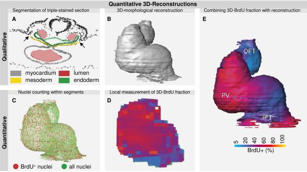
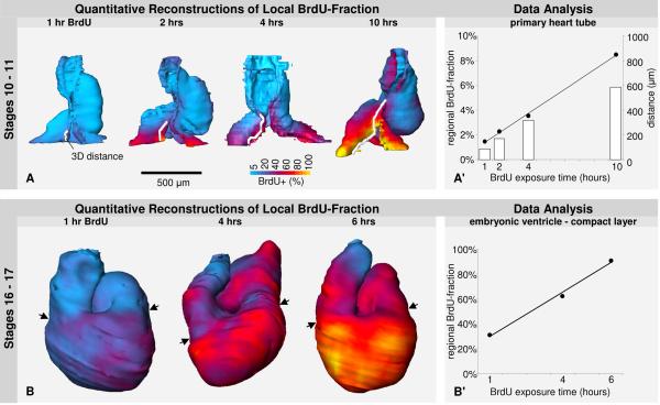
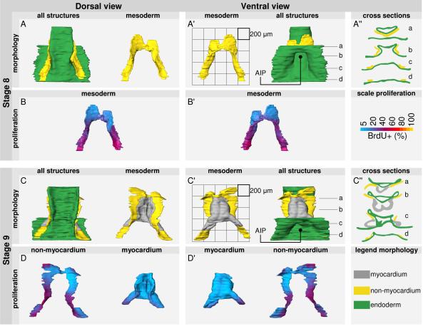
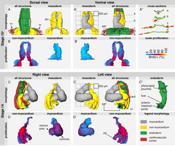
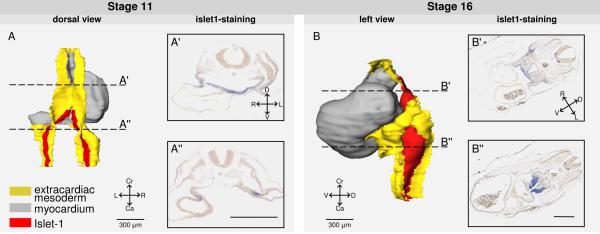
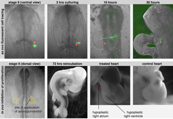
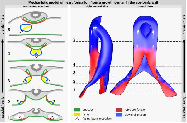
Recent studies have shown that the primary heart tube continues to grow by addition of cells from the coelomic wall. This growth occurs concomitantly with embryonic folding and formation of the coelomic cavity, making early heart formation morphologically complex. A scarcity of data on localized growth parameters further hampers the understanding of cardiac growth. Therefore, we investigated local proliferation during early heart formation. Firstly, we determined the cell cycle length of primary myocardium of the early heart tube to be 5.5 days, showing that this myocardium is nonproliferating and implying that initial heart formation occurs solely by addition of cells. In line with this, we show that the heart tube rapidly lengthens at its inflow by differentiation of recently divided precursor cells. To track the origin of these cells, we made quantitative 3D reconstructions of proliferation in the forming heart tube and the mesoderm of its flanking coelomic walls. These reconstructions show a single, albeit bilateral, center of rapid proliferation in the caudomedial pericardial back wall. This center expresses Islet1. Cell tracing showed that cells from this caudal growth center, besides feeding into the venous pole of the heart, also move cranially via the dorsal pericardial mesoderm and differentiate into myocardium at the arterial pole. Inhibition of caudal proliferation impairs the formation of both the atria and the right ventricle. These data show how a proliferating growth center in the caudal coelomic wall elongates the heart tube at both its venous and arterial pole, providing a morphological mechanism for early heart formation.


Figures

Figure 1
Figure 1
Panel A shows a representative image of the Sytox-green channel of a triple-stained section. Overlaying the image are the segmented myocardium (grey), splanchnic mesoderm (yellow), the endoderm (green) and the cardiovascular lumen (red). Arrows indicate the borders between intra and extra-embryonic mesoderm. BrdU-positive and all nuclei were automatically identified within the myocardium and selected splanchnic mesoderm. Panel C shows nuclei of the myocardium in their 3D-context. To facilitate interpretation, and allow reliable estimations, nuclei were counted and BrdU-fractions were determined in sample-cubes, as shown in panel D. This information is then projected on the morphological reconstruction (panel B) to give a quantitative 3D-reconstruction of local proliferation (panel E). (Displayed reconstruction is the myocardium of a stage 12 chicken embryo, after 4 hours of BrdU-exposure.6)
Figure 2
Figure 2
Panels A and B show quantitative 3D-reconstructions of local BrdU+-fractions of embryos that were increasingly exposed to BrdU. The nuclear BrdU-fractions are presented using a color-bar, ranging from blue (0%) to yellow (100%). The length of expanding BrdU-positive zone along the left dorsal mesocardium was measured and indicated in white. The relation of this expansion to the BrdU exposure-time is shown in panel C. To calculate cell-cycle times we exploited the linear relation between BrdU-fractions (FB) and BrdU-exposure time (TB). This relation follows the equation: FB = TS/TG + 1/TG × TB, with TS and TG representing the length of the S-phase and the cell cycle, respectively. The inverse of the slope of this linear relation equals the cell-cycle length. For calculations of the primary heart tube we counted BrdU+-fractions of the reconstructions in panel B, with exclusion of the expanding zone of BrdU-labeled cells. For calculations of ventricular myocardium, we counted BrdU+-fractions in the compact layer at the outer curvature of the reconstructions in panel B, between the distal ventricular groove (left arrow) and the atrioventricular canal (right arrow). Panel D shows these linear relations of both the ventricular and the primary myocardium (hatched line).
Figure 3
Figure 3
shows morphology and proliferation (as defined by the BrdU+-fraction after 1 hour of exposure) of the heart region of stage 8 (rows A and B) and stage 9 (rows C and D) chicken embryos. Myocardium is shown in grey, non-myocardial mesoderm in yellow and endoderm in green. In A' and C' a scale grid of (1000 μm)2 is shown. Also, in A' and C' locations are indicated of cross-sections that are shown in A” and C”, respectively. (AIP: anterior intestinal portal)
Figure 4
Figure 4
shows morphology and proliferation (as defined by the BrdU+-fraction after 1 hour of exposure) of the heart region of stage 10+ (rows A and B) and stage 14 (rows C and D) chicken embryos. The myocardium is shown in grey, non-myocardial mesoderm in yellow, endoderm in green, and cardiovascular lumen in red. In A' and C' a scale grid of (1000 μm)2 is shown. Also, in A' locations are indicated of cross-sections that are shown in A”. Panel C” shows the reconstructed endoderm. (AIP: anterior intestinal portal, card: cardinal vein, dm: dorsal mesocardium, PE: proepicardium, vv: vitelline veins, △: outer mesoderm that covers the vitelline veins, ▲: inner mesoderm that covers the anterior intestinal portal, hatched arrows: pericardioperitoneal canals)
Figure 5
Figure 5
shows reconstructions of extension of the expression of Islet1-mRNA in red. Reconstructed are the myocardium (grey) and non-myocardial mesoderm (yellow). Panel A shows a dorsal view of a stage 11 embryo. Section A' shows expression of Islet1 in the endoderm. Section A” shows expression in the pericardioperitoneal canals. Panel B shows a left view of a stage 16 embryo. Section B' shows expression in the pericardial back wall. Section B” shows expression in the pericardioperitoneal canals. (Ca: caudal, Cr: cranial, D: dorsal, L: left, R: right, V: ventral) Scale bars: 300 μm.
Figure 6
Figure 6
Panel A shows the movement of fluorescently labeled cells from the caudal splanchnic mesoderm of a stage 9 embryo. The inner mesoderm is labeled with DiO (green) and the outer mesoderm is labeled with DiI (red). For a spatial appreciation of the inner and outer mesoderm refer to Figure 4 (indicated with ▲ and △, respectively). With culturing the outer mesoderm can be seen to be incorporated into the inflow of the heart, while the inner mesoderm moves, via the dorsal pericardial wall, into the outflow of the heart. Panel B shows the effect of local inhibition of proliferation. At the right side of the embryo, a focus in the caudal and inner mesoderm was exposed to Aminopurvanolol, dissolved with DiI. After reincubation 9 out of 16 treated embryos suffered from hypoplasia of both the right ventricle and the right atrium. Of the other embryos, 2 deceased before analysis, 1 showed only outflow-malformations, and 4 seemed to be unaffected.
Figure 7
Figure 7
illustrates heart formation from the proliferating growth-center in the dorsal pericardial wall. The left column shows transverse sections, ranging from cranial in an old embryo to caudal in a young embryo. Firstly, outer mesoderm luminizes and stops to proliferate. Next, it bends inwards and fuses to form the ventral wall of the heart tube (2-4). The inner mesoderm keeps proliferating and forms the pericardial back wall and its connection with the heart tube (5). These sections were transformed into the model that is shown on the right. The model shows that expansion from the caudal growth-center leads to a radial addition of medially located mesoderm to the heart tube. After regression of the dorsal mesocardium, addition to the arterial pole occurs via the pericardial back wall.

References

    1. DeHaan RL. Development of form in the embryonic heart. An experimental approach. Circulation. 1967;35:821–833. - PubMed
    1. Moorman AFM, Christoffels VM. Cardiac chamber formation: development, genes and evolution. Physiol Rev. 2003;83:1223–1267. - PubMed
    1. Hoffman JI, Kaplan S, Liberthson RR. Prevalence of congenital heart disease. Am Heart J. 2004;147:425–439. - PubMed
    1. Sissman J. Cell multiplication rates during development of the primitive cardiac tube in the chick embryo. Nature. 1966;210:504–507. - PubMed
    1. Stalsberg H. Regional mitotic activity in the precardiac mesoderm and differentiating heart tube in the chick embryo. Dev Biol. 1969;20:18–45. - PubMed

Publication types

MeSH terms

Substances