. 2009 Jul;41(1):107-13.
doi: 10.1165/rcmb.2008-0381OC. Epub 2008 Dec 4.

NAD(P)H quinone oxidoreductase 1 is essential for ozone-induced oxidative stress in mice and humans

Affiliations

NAD(P)H quinone oxidoreductase 1 is essential for ozone-induced oxidative stress in mice and humans

Judith A Voynow et al. Am J Respir Cell Mol Biol. 2009 Jul.

Erratum in

Abstract

One host susceptibility factor for ozone identified in epidemiologic studies is NAD(P)H quinone oxidoreductase 1 (NQO1). We hypothesized that after ozone exposure, NQO1 is required to increase 8-isoprostane (also known as F(2)-isoprostane) production, a recognized marker of ozone-induced oxidative stress, and to enhance airway inflammation and hyperresponsiveness. In this report, we demonstrate that in contrast to wild-type mice, NQO1-null mice are resistant to ozone and have blunted responses, including decreased production of F(2)-isoprostane and keratinocyte chemokine, decreased airway inflammation, and diminished airway hyperresponsiveness. Importantly, these results in mice correlate with in vitro findings in humans. In primary human airway epithelial cells, inhibition of NQO1 by dicumarol blocks ozone-induced F(2)-isoprostane production and IL-8 gene expression. Together, these results demonstrate that NQO1 modulates cellular redox status and influences the biologic and physiologic effects of ozone.


Figures

<b>Figure 1.</b>
Figure 1.
Airway hyperresponsiveness in C57BL/6 wild-type and NAD(P)H quinone oxidoreductase 1 (NQO1)-null mice after filtered air (FA) or ozone (OZ) exposure. Twenty-four hours after OZ or FA exposure, mice were anesthetized for invasive measures of total pulmonary resistance in response to methacholine by impulse oscillometry with the flexivent ventilator (n = 5 animals/group; mean ± SEM; *significantly different from corresponding air control, P < 0.05; #significantly different from WT-ozone exposed animals, P < 0.05).
<b>Figure 2.</b>
Figure 2.
NQO1 Western analyses in C57BL/6 wild-type and NQO1-null mice after FA or OZ exposure. C57BL/6 mice and congenic strain, NQO1-null mice were exposed to OZ (1 ppm, 3 h) or FA. Immediately after exposure (0 h) and 6, 12, 24, and 48 hours after OZ or FA exposure, lungs were harvested for NQO1 protein expression. (A) Western analyses were performed for NQO1. Total lung protein (50 μg) was separated by 12% SDS-PAGE, transferred to a nitrocellulose membrane, and incubated with a polyclonal goat-anti NQO1 antibody (1:1,000), followed by HRP-conjugated anti-goat IgG (1:5,000). Antigen–antibody complexes were visualized by chemiluminescence with ECL plus and autoradiography. Western analyses for β-actin were performed to confirm equivalent protein loading. (B) The graph was made from two separate autoradiographs; the 24-hour time point data, from a separate autoradiograph, was added to the autoradiograph which included the data from the other time points. NQO1 band densities were quantitated and expressed relative to β-actin band densities, and then normalized to the corresponding FA-exposed animals. Dashed line represents 0-time control-FA levels (mean ± SEM, n = 5 animals/group).
<b>Figure 3.</b>
Figure 3.
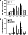
Bronchoalveolar lavage total and neutrophil cell counts in C57BL/6 wild-type and NQO1-null mice after FA or OZ exposure. Immediately after FA or OZ exposure (0 h), and 6, 12, 24, and 48 hours after exposure, bronchoalveolar lavage (BAL) was performed as described in Materials and Methods. (A) Total cell counts and (B) neutrophil counts were determined in wild-type and NQO1-null mice exposed to FA or OZ (n = 5 animals/group). Results are expressed as mean ± SEM. *OZ-exposed significantly different from FA-exposed, P < 0.05; #OZ-exposed NQO1-null mice are significantly different from wild-type OZ-exposed mice, P < 0.05.
<b>Figure 4.</b>
Figure 4.
Keratinocyte chemoattractant (KC) levels in BAL by ELISA in C57BL/6 wild-type and NQO1-null mice after OZ exposure. C57BL/6 mice and congenic strain, NQO1-null mice were exposed to ozone (1 ppm, 3 h) or FA. Immediately after OZ exposure (0 h), and 6, 12, 24, and 48 hours after exposure, BAL was performed as described in Materials and Methods. BAL was used for ELISA assays to quantitate the neutrophil chemotactic chemokine, KC (n = 5 animals/group), according to the manufacturer's instructions. Results are expressed as mean ± SEM. *OZ-exposed significantly different from FA-exposed, P < 0.05; #wild-type OZ-exposed mice significantly different from OZ-exposed NQO1-null mice, P < 0.05.
<b>Figure 5.</b>
Figure 5.
F2-isoprostane levels in BAL by EIA in C57BL/6 wild-type and NQO1-null mice after OZ exposure. BAL was performed at 0, 6, 12, and 48 hours after ozone (OZ) or filtered air (FA), and then analyzed for F2-isoprostane by EIA according to the manufacturer's instructions. BAL KC quantitation is presented as pg/ml (mean ± SEM, n = 5 animals/group).
<b>Figure 6.</b>
Figure 6.
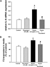
IL-8 gene expression by real-time RT-PCR and F2-isoprostane quantitation by EIA in cultured primary normal human bronchial epithelial cells after OZ exposure. NHBE cells cultured at air–liquid interface (ALI) were preincubated and coincubated with NQO1 inhibitor, dicumarol (10 nM, 1 h), or control vehicle at the apical and basolateral compartments, and then exposed to OZ (0.4 ppm, 5 h) or FA with media ± dicumarol only in the basolateral compartments. At the end of the exposure period, total RNA was collected using Trizol reagent, and medium was collected for F2-isoprostane measurements by EIA. As per the manufacturer's instructions, BHT was added to the medium to a final concentration of 0.005%. (A) Real-time RT-PCR analysis of IL-8 mRNA expression was performed as described in Materials and Methods (n = 6). (B) F2-isoprostane levels in the cell culture medium were quantitated and expressed as a percentage of the control for each experiment (n = 11–12; two to four separate experiments, mean ± SEM are shown). *OZ-exposed cells significantly greater than control, P < 0.05; #OZ+dicumarol–exposed cells were significantly different from OZ-exposed cells, P < 0.05.

Comment in

  • Findings of Research Misconduct.
    [No authors listed] [No authors listed] Fed Regist. 2019 Nov 7;84(216):60097-60098. Fed Regist. 2019. PMID: 37547121 Free PMC article. No abstract available.

Similar articles

Cited by

References

    1. American Thoracic Society. Health effects of outdoor air pollution. Part 2. Committee of the Environmental and Occupational Health Assembly of the American Thoracic Society. Am J Respir Crit Care Med 1996;153:477–498. - PubMed
    1. Peden DB. The epidemiology and genetics of asthma risk associated with air pollution. J Allergy Clin Immunol 2005;115:213–219. (quiz 220). - PubMed
    1. Foster W. Effects of oxidants. In: Swift D, Foster W, editors. Air pollutants and the respiratory tract. New York: Marcel Dekker; 1999. pp. 147–179.
    1. Trasande L, Thurston GD. The role of air pollution in asthma and other pediatric morbidities. J Allergy Clin Immunol 2005;115:689–699. - PubMed
    1. Gent JF, Triche EW, Holford TR, Belanger K, Bracken MB, Beckett WS, Leaderer BP. Association of low-level ozone and fine particles with respiratory symptoms in children with asthma. JAMA 2003;290:1859–1867. - PubMed

Publication types

MeSH terms