. 2009 Feb;37(2):e12.
doi: 10.1093/nar/gkn954. Epub 2008 Dec 5.

RNA cytosine methylation analysis by bisulfite sequencing

Affiliations

RNA cytosine methylation analysis by bisulfite sequencing

Matthias Schaefer et al. Nucleic Acids Res. 2009 Feb.

Abstract

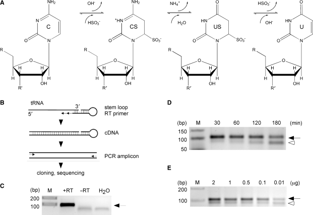
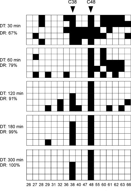
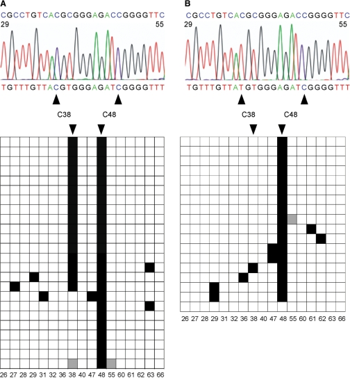
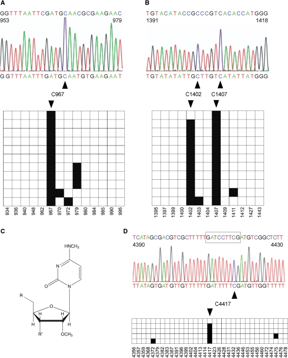
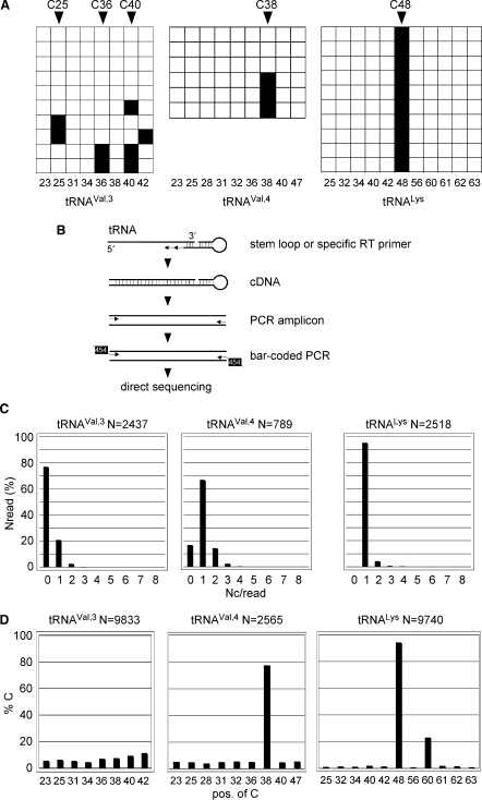
Covalent modifications of nucleic acids play an important role in regulating their functions. Among these modifications, (cytosine-5) DNA methylation is best known for its role in the epigenetic regulation of gene expression. Post-transcriptional RNA modification is a characteristic feature of noncoding RNAs, and has been described for rRNAs, tRNAs and miRNAs. (Cytosine-5) RNA methylation has been detected in stable and long-lived RNA molecules, but its function is still unclear, mainly due to technical limitations. In order to facilitate the analysis of RNA methylation patterns we have established a protocol for the chemical deamination of cytosines in RNA, followed by PCR-based amplification of cDNA and DNA sequencing. Using tRNAs and rRNAs as examples we show that cytosine methylation can be reproducibly and quantitatively detected by bisulfite sequencing. The combination of this method with deep sequencing allowed the analysis of a large number of RNA molecules. These results establish a versatile method for the identification and characterization of RNA methylation patterns, which will be useful for defining the biological function of RNA methylation.


Figures

Figure 1.
Figure 1.
Establishment of RNA bisulfite sequencing. (A) Schematic diagram of the bisulfite conversion reaction. C, cytidine; CS cytidine sulfonate; US, uridine sulfonate; U, uridine. (B) Outline of the basic strategy to analyze tRNA for m5C methylation. Bisulfite-treated tRNAs are reverse transcribed using a tRNA 3′-sequence-specific stem–loop primer, amplified with primers binding only to deaminated sequences at the 5′ end, followed by standard cloning and sequencing. (C) As an example, in vitro transcribed tRNAAsp served as template for cDNA synthesis. RT, reverse transcriptase; arrow, tRNA amplicon. (D) Increasing deamination times lead to degradation of tRNAAsp. Equal amounts of cellular RNA (1 μg) were subjected to deamination for the time indicated, followed by cDNA synthesis and PCR amplification. (E) Dilution series of total RNA subjected to bisulfite treatment, followed by cDNA synthesis and PCR amplification of tRNAAsp. Ten nanograms of cellular RNA are sufficient to amplify bisulfite-treated tRNAAsp (32 amplification cycles). Arrow, tRNA amplicon; open arrowhead, aberrant PCR amplicon.
Figure 2.
Figure 2.
In vivo RNA bisulfite sequencing of tRNAAsp from total RNA (1 μg) after various deamination times. Five clones were sequenced for each time point and deamination rates (DR) were calculated based on the number of all cytosines (minus known m5C residues at positions C38 and C48). Black boxes indicate methylated cytosine residues, white boxes indicate unmethylated cytosine residues. Each PCR amplicon queried 17 cytosines in tRNAAsp (C = 17). Numbers below boxes indicate the cytosine positions in the primary RNA sequence.
Figure 3.
Figure 3.
Validation of RNA bisulfite sequencing by the detection of tRNAAsp methylation changes in Dnmt2 mutant tissues. (A) Bisulfite sequencing analysis of tRNAAsp from wild-type D. melanogaster embryos. A representative sequencing trace showing the deaminated tRNAAsp sequence (nucleotides 29–55 containing nine cytosines) with the nondeaminated sequence for comparison (above). The diagram shows the methylation status of individual cytosines (horizontal, C=17) in several independent clones (vertical, N = 24). Black boxes indicate methylated cytosine residues, white boxes indicate unmethylated cytosine residues, gray boxes indicate cytosine residues with unknown methylation status (due to bad sequence reads). Numbers below boxes indicate the cytosine positions in the primary RNA sequence. The deamination rate was calculated to be 98%. C38 (methylated by the Dnmt2 methyltransferase) and C48 (methylated by the Trm4 methyltransferase) were methylated in 70% and 100% of the sequences, respectively. (B) Bisulfite sequencing analysis of tRNAAsp from Dnmt2 mutant D. melanogaster embryos. The deamination rate was calculated to be 97%. C38 and C48 were methylated in 6% and 94% of the sequences (N = 18), respectively.
Figure 4.
Figure 4.
RNA bisulfite sequencing detects cytosine methylation in ribosomal RNAs. Black boxes indicate methylated cytosine residues, white boxes indicate unmethylated cytosine residues. (A) A representative sequencing trace showing the deaminated 16S rRNA from E. coli (nucleotides 953–979 containing five cytosines) with the nondeaminated sequence for comparison (above). The diagram shows the methylation status of individual cytosines (horizontal, C = 14) in several independent clones (vertical, N = 11). The deamination rate was calculated to be 96%. C967 (methylated by the RsmB/Fmu methyltransferase) was methylated in 100% of the sequences. (B) Parallel analysis of nucleotides 1391–1418 from E. coli 16S rRNA. C1407 (methylated by the RsmF/YebU methyltransferase) was methylated in 100% of the sequences. Interestingly, C1402 containing m4mC, N4,2′-O-dimethylcytidine, was scored as methylated in 88% of the sequences. The diagram shows the methylation status of individual cytosines (horizontal, C = 13) in several independent clones (vertical, N = 11). The deamination rate was calculated to be 98%. (C) Chemical structure of N4,2′-O-dimethylcytidine which can be detected by RNA bisulfite sequencing at position C1402 in 16S rRNA. (D) Precision mapping of m5C in human 28S rRNA. A representative sequencing trace showing the deaminated 28S rRNA from the human HCT116 cell line (nucleotides 4390–4430) with the nondeaminated sequence for comparison (above). The boxed sequence highlights the region to which m5C was localized previously (20). The diagram shows the methylation status of individual cytosines (horizontal, C = 33) in five independent clones. The deamination rate was calculated to be 99%. Bisulfite sequencing uncovers the exact position of m5C at nucleotide 4417. Numbers below boxes indicate the cytosine positions in the primary RNA sequence.
Figure 5.
Figure 5.
Deep sequencing of tRNA methylation patterns. (A) Conventional sequencing results. Bisulfite sequencing analysis of tRNAs from wild-type D. melanogaster embryos. The diagram shows the methylation status of individual cytosines (horizontal squares) in several independent clones (vertical squares) in tRNAVal,3, tRNAval,4 and tRNALys. Black boxes indicate methylated cytosine residues, white boxes indicate unmethylated cytosine residues. Numbers below boxes show cytosine position in primary RNA sequence. (B) Outline of the strategy to analyze tRNA for m5C methylation using 454 technology. Bisulfite-treated tRNAs are reverse transcribed using a stem–loop primer or a specific tRNA primer (tRNAVal), followed by PCR amplification step 1 with primers binding only to deaminated sequences at the 5′ end and step 2 with primers introducing the bar-coded 454 sequence. PCR amplicons were sequenced directly. (C) 454 deep-sequencing results. Distribution of nondeaminated cytosines per read (Nc/read) for the illustration of pattern complexity and deamination efficiency. (D) Bisulfite sequencing analysis of tRNAs from wild-type D. melanogaster embryos. Fractions of nondeaminated cytosines are plotted against the position of the cytosine in individual tRNA amplicons, thus revealing the pattern of m5C modification in individual tRNAs.

References

    1. Suzuki MM, Bird A. DNA methylation landscapes: provocative insights from epigenomics. Nat. Rev. Genet. 2008;9:465–476. - PubMed
    1. Rozenski J, Crain PF, McCloskey JA. The RNA Modification Database: 1999 update. Nucleic Acids Res. 1999;27:196–197. - PMC - PubMed
    1. Rein T, DePamphilis ML, Zorbas H. Identifying 5-methylcytosine and related modifications in DNA genomes. Nucleic Acids Res. 1998;26:2255–2264. - PMC - PubMed
    1. Thomassin H, Oakeley EJ, Grange T. Identification of 5-methylcytosine in complex genomes. Methods. 1999;19:465–475. - PubMed
    1. Frommer M, McDonald LE, Millar DS, Collis CM, Watt F, Grigg GW, Molloy PL, Paul CL. A genomic sequencing protocol that yields a positive display of 5-methylcytosine residues in individual DNA strands. Proc. Natl Acad. Sci. USA. 1992;89:1827–1831. - PMC - PubMed

Publication types