. 2009 Feb;191(4):1132-42.
doi: 10.1128/JB.01359-07. Epub 2008 Dec 5.

Mycobacterium avium genes MAV_5138 and MAV_3679 are transcriptional regulators that play a role in invasion of epithelial cells, in part by their regulation of CipA, a putative surface protein interacting with host cell signaling pathways

Affiliations

Mycobacterium avium genes MAV_5138 and MAV_3679 are transcriptional regulators that play a role in invasion of epithelial cells, in part by their regulation of CipA, a putative surface protein interacting with host cell signaling pathways

Melanie J Harriff et al. J Bacteriol. 2009 Feb.

Abstract

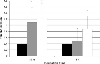
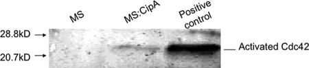
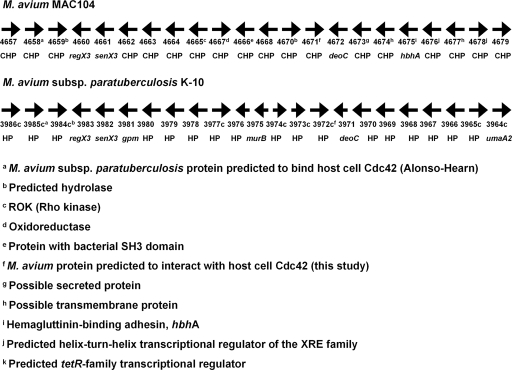
The Mycobacterium avium complex (MAC) is an important group of opportunistic pathogens for birds, cattle, swine, and immunosuppressed humans. Although invasion of epithelial cells lining the intestine is the chief point of entry for these organisms, little is known about the mechanisms by which members of the MAC are taken up by these cells. Studies with M. avium have shown that cytoskeletal rearrangement via activation of the small G-protein Cdc42 is involved and that this activation is regulated in part by the M. avium fadD2 gene. The fadD2 gene indirectly regulates a number of genes upon exposure to HEp-2 cells, including transcriptional regulators, membrane proteins, and secreted proteins. Overexpression of two fadD2-associated regulators (MAV_5138 and MAV_3679) led to increased invasion of HEp-2 cells, as well as altered expression of other genes. The protein product of one of the regulated genes, named CipA, has domains that resemble the PXXP motif of human Piccolo proteins, which bind SH3 domains in proteins involved in the scaffold complex formed during cytoskeletal rearrangement. Although CipA was not detected in the cytoplasm of HEp-2 cells exposed to M. avium, the recombinant protein was shown to be potentially expressed on the surface of Mycobacterium smegmatis incubated with HEp-2 cells and, possibly, to interact with human Cdc42. The interaction was then confirmed by showing that CipA activates Cdc42. These results suggest that members of the M. avium complex have a novel mechanism for activating cytoskeletal rearrangement, prompting uptake by host epithelial cells, and that this mechanism is regulated in part by fadD2, MAV_5138, and MAV_3679.


Figures

FIG. 1.
FIG. 1.
Real-time PCR results comparing the fold change in gene expression upon 15 min of exposure to HEp-2 cells between the MAC109 and MAC109 ΔfadD2 strains. The y axis represents the fold change between broth grown bacteria and bacteria incubated for 15 min with HEp-2 cells, using the 16S rDNA as an internal control. An asterisk (*) indicates significant differences between the two strains, based on three independent experiments (P < 0.05). Columns: ▪, MAC109; ▪, MAC109 ΔfadD2.
FIG. 2.
FIG. 2.
Percent invasion of HEp-2 cells by strains of M. avium overexpressing MAV_3679 or MAV_5138 after a 30-min or 1-h incubation period. Black bars represent MAC109, gray bars represent MAC109/pMH6, and white bars represent MAC109/pMH7. Both recombinant strains have significantly higher invasion than MAC109 after 30 min, and MAC109/pMH7 also has a significantly higher invasion after 1 h. An asterisk (*) indicates significant differences from the baseline, based on three independent experiments (P < 0.05). Columns: ▪, MAC109; ▪, MAC109:MAV_5138; □, MAC109:MAV_3679.
FIG. 3.
FIG. 3.
Fold change in expression of genes in M. avium overexpressing MAV_3679 (A) or MAV_5138 (B) compared to wild-type MAC109 grown in culture medium. The results shown are the average and standard error of three experiments.
FIG. 4.
FIG. 4.
Domains/regions present in the CipA amino acid sequence.
FIG. 5.
FIG. 5.
CipA expressed as a fusion protein with the B. pertussis adenylate cyclase gene is not detected in the cytosolic or insoluble fractions of HEp-2 cells infected with M. avium. Proteins were extracted from broth-grown M. avium or from HEp-2 cells infected with strains of M. avium. An α-CyaA antibody and agarose-conjugate α-IgG beads were used to precipitate the CipA:CyaA fusion protein from the lysates. (A) Protein gel stained with Coomassie blue showing the expression of the 80-kDa CipA:CyaA fusion protein (arrow) in broth-grown M. avium. Lane 1, MAC104; lane 2, MAC104/pMH5. (B) Western blot showing proteins immunoprecipitated from HEp-2 cells infected with strains of M. avium. Lane 3, soluble lysate fraction incubated with MAC104; lane 4, soluble lysate fraction incubated with MAC104/pMH5; lane 5, Insoluble lysate fraction incubated with MAC104; lane 6, insoluble lysate fraction incubated with MAC104/pMH5.
FIG. 6.
FIG. 6.
(A and B) HEp-2 cells exposed to M. smegmatis expressing CipA fused to GFP or expressing GFP alone. There is a polar accumulation of GFP/CipA. (A) Representative images of individual bacterial cells, expressing the fusion protein, in contact with cytochalasin D-treated HEp-2 cells. (B) Representative images of individual bacterial cells expressing GFP only, in contact with cytochalasin D-treated HEp-2 cells. (C to E) Cells infected with either strain of M. smegmatis, or fluroescein-labeled M. avium were incubated with α-Cdc42 antibodies and a Texas Red-conjugated secondary antibody. (C) Cells infected with M. avium for 15 min; (D) cells infected with M. avium for 30 min; (E) cells infected with M. smegmatis for 1 h. The figure shows that, while M. avium appears to interact with Cdc42 on the cell, M. smegmatis does not.
FIG. 7.
FIG. 7.
Western blot showing that CipA is capable of activating host cell Cdc42. HEp-2 cells were incubated either with M. smegmatis wild-type or M. smegmatis expressing CipA in the membrane. HEp-2 cells were lysed and positive control were treated with GTPγδ to activate Cdc42. Activated Cdc42 was captured as described in Materials and Methods. Proteins were eluted and resolved by SDS-PAGE, transferred to a membrane, and probed with anti-Cdc42 antibody. Lane 1, M. smegmatis not expressing CipA; lane 2, positive control; lane 3, M. smegmatis expressing CipA.
FIG. 8.
FIG. 8.
M. avium (numbers represent MAV annotation) and M. avium subsp. paratuberculosis (numbers represent MAP annotation) chromosomal regions encompassing the CipA gene discussed in the present study. CHP, conserved hypothetical protein; HP, hypothetical protein.

References

    1. Alonso-Hearn, M., D. Patel, L. Danelishvili, L. Meunier-Goddik, and L. E. Bermudez. 2008. The Mycobacterium avium subsp. paratuberculosis MAP3464 gene encodes an oxidoreductase involved in invasion of bovine epithelial cells through the activation of host cell Cdc42. Infect. Immun. 76170-178. - PMC - PubMed
    1. Ando, M., Y. C. Manabe, P. J. Converse, E. Miyazaki, R. Harrison, J. R. Murphy, and W. R. Bishai. 2003. Characterization of the role of the divalent metal ion-dependent transcriptional repressor MntR in the virulence of Staphylococcus aureus. Infect. Immun. 712584-2590. - PMC - PubMed
    1. Bach, H., J. Sun, Z. Hmama, and Y. Av-Gay. 2006. Mycobacterium avium subsp. paratuberculosis PtpA is an endogenous tyrosine phosphatase secreted during infection. Infect. Immun. 746540-6546. - PMC - PubMed
    1. Barber, C. E., J. L. Tang, J. X. Feng, M. Q. Pan, T. J. Wilson, H. Slater, J. M. Dow, P. Williams, and M. J. Daniels. 1997. A novel regulatory system required for pathogenicity of Xanthomonas campestris is mediated by a small diffusible signal molecule. Mol. Microbiol. 24555-566. - PubMed
    1. Bendtsen, J. D., H. Nielsen, G. von Heijne, and S. Brunak. 2004. Improved prediction of signal peptides: SignalP 3.0. J. Mol. Biol. 340783-795. - PubMed

Publication types

LinkOut - more resources