. 2008 Dec 16;105(50):19587-94.
doi: 10.1073/pnas.0809577105. Epub 2008 Dec 5.

Light-arousal and circadian photoreception circuits intersect at the large PDF cells of the Drosophila brain

Affiliations

Light-arousal and circadian photoreception circuits intersect at the large PDF cells of the Drosophila brain

Yuhua Shang et al. Proc Natl Acad Sci U S A. .

Abstract

The neural circuits that regulate sleep and arousal as well as their integration with circadian circuits remain unclear, especially in Drosophila. This issue intersects with that of photoreception, because light is both an arousal signal in diurnal animals and an entraining signal for the circadian clock. To identify neurons and circuits relevant to light-mediated arousal as well as circadian phase-shifting, we developed genetic techniques that link behavior to single cell-type resolution within the Drosophila central brain. We focused on the unknown function of the 10 PDF-containing large ventral lateral neurons (l-LNvs) of the Drosophila circadian brain network and show here that these cells function in light-dependent arousal. They also are important for phase shifting in the late-night (dawn), indicating that the circadian photoresponse is a network property and therefore non-cell-autonomous. The data further indicate that the circuits underlying light-induced arousal and circadian photoentrainment intersect at the l-LNvs and then segregate.


Conflict of interest statement

The authors declare no conflict of interest.

Figures
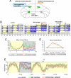
Fig. 1.
Fig. 1.
l-LNvs are part of the peptidergic arousal pathways in Drosophila. (A) Genetic scheme for the 2 mutants is shown. (B and C) Constitutive firing of PHM+ peptidergic cells dramatically increased activity at night in LD conditions. LD activity/rest pattern was monitored from groups of flies with 3 different genotypes: control 929:dORKΔNC1 (n = 14), PHM+ 929:NaChBac (n = 20), and PHM+ PDF 929:NaChBac;pdfGal80 (n = 29). Experiments were repeated >3 times. dORKΔNC1GFP is a nonconducting K+ channel and was used as control. UAS-NaChBacGFP flies showed no abnormal nocturnal activity or sleep like that seen in 929:NaChBacGFP flies (data not shown). (B) Double-plotted actograms are shown. Light phase is highlighted in yellow and dark phase is highlighted in gray. Red brackets indicate increase of nighttime activity in mutant flies (Left) and black arrows (Center and Right) indicate rescue of early night behavior by pdfGal80. (C) The daytime and nighttime activity of the genotypes are compared. (D) Heat-induced acute firing of peptidergic cells and l-LNvs caused a transient increase of locomotor activity and a long-term suppression of sleep homeostasis. The rest–activity behavior was monitored from groups of flies with 3 different genotypes: control (n = 23), 929:dTrpA1 (n = 20), and 929:dTrpA1;pdfGal80 (n = 19). (E) Constant light abolished the arousal-promoting effect of hyperexciting l-LNvs and other peptidergic cells. LD and LL sleep was monitored from groups of flies with 3 different genotypes: control 929:dORKΔNC1 (n = 20), PHM+ 929:NaChBac (n = 17), and PHM+ PDF 929:NaChBac;pdfGal80 (n = 16).
Fig. 2.
Fig. 2.
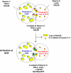
A subset of l-LNvs can be labeled by the intersectional technique. The flies carries 5 transgenes: promoter A-Gal4, UAS-NaChBacGFP, tub>GAL80>, promoter B-LexA, and LexAop-Flp. GAL80 is flanked by 2 FRT sites (>) and is ubiquitously expressed by the tubulin promoter (yellow). “Neuron 1” are groups of neurons expressing GAL4 proteins (blue). “Neuron 2” are groups of neurons expressing LexA transcription factors (red). “Neuron 1” and “Neuron 3” intersect at “Neuron 2,” which express both GAL4 and LexA proteins (blue and red). GAL80 proteins (yellow) suppress the GAL4-induced expression of genes under UAS control with no effect on LexA-induced expression. FLP causes the Gal80 coding region to be removed in a subset of neurons 2 and 3. As a result, a subset of neuron 3 will lose Gal80 coding sequence, thereby activating GAL4-driven UAS-NaChBacGFP transcription (green).
Fig. 3.
Fig. 3.
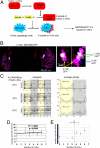
Stimulation of l-LNvs is sufficient to promote arousal at night. (A) The combination of GAL4-UAS/GAL80, LexA-LexAop expression system with the FLP/FRT system in the same fly. The l-LNv> NaChBacGFP fly carries 5 transgenes: c929Gal4, UAS-NaChBacGFP, tub>GAL80>, pdfLexA, and LexAopFlp (for details, see Materials and Methods). (B) A small subset of l-LNvs can be labeled by the intersectional technique. A brain from a fly w; c929Gal4,UAS-NaChBacGFP/tub>GAL80>; pdfLexA,LexAopFLP/+ was fixed and stained with antibody against GFP (green) and PDF (magenta). The higher magnification (on the right) shows 3 of 10 l-LNvs in this fly expressing NaChBacGFP channel (white). No GFP staining can be detected outside the l-LNvs. (C) Examples of activity/rest pattern in LD and genotype of l-LNvs from 3 individual flies. (D and E) Increasing the excitability of l-LNvs at night directly promotes activity. Twenty-eight flies expressing no GFP outside the l-LNvs were divided into subgroups based on the number of l-LNvs expressing NaChBacGFP. The mean daytime (D) or nighttime (E) activity was then calculated within each group. The correlation between the number of l-LNvs labeled and the mean activity during daytime or nighttime is plotted separately. The number of l-LNvs expressing NaChBacGFP channel is highly correlated with the nighttime activity, without affecting daytime activity. A threshold effect was observed, i.e., exciting 4 or more l-LNvs is sufficient to induce nighttime hyperactivity (E). **, P < 0.01.
Fig. 4.
Fig. 4.
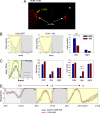
l-LNvs are part of the light-mediated arousal pathway. (A) Ectopic expression of hid, a cell death gene, lead to loss of l-LNvs. Brains from l-LNv> hid flies were fixed and stained with PDF antibody. This fly had only 2 l-LNvs in the left hemisphere and no l-LNvs in the right hemisphere (yellow arrow). The PDF signal is largely reduced in the left optical lobe and almost completely lost in the right optical lobe (green arrow). s-LNvs appear normal (white arrows). (B) Loss of l-LNvs significantly reduced the daytime activity in female flies. The actogram, average activity, and mean duration of wakefulness for control (c929Gal4,UASGFP/+;pdfLexA,LexAopFLP/+, n = 9) and l-LNv> hid flies (n = 18) are shown. ***, P < 0.001. (C) Loss of l-LNvs significantly increases the daytime sleep in female flies. Sleep graph, total sleep, and maximum sleep for the 2 genotypes are shown. ****, P < 0.0001. In B and C, blue box is control and red box is flies with fewer l-LNvs. Female flies expressing hid (n = 32 for control and n = 52 for hid flies total) consistently showed less activity and more sleep during the day. (D) l-LNv>hid flies have no arousal/sleep phenotype in the first subjective day of DD compared with that of LD, whereas the arousal/sleep phenotype is more obvious in LL condition. Control flies (UAS>hid/+;pdfLexA,LexAopFLP/+, n = 34) and l-LNv>hid flies (n = 10) were entrained in LD for 5 days, released into DD for 2 days, reentrained in LD for 2 days, and released into LL. The sleep graph in DD, LD, and LL for both genotypes is shown.
Fig. 5.
Fig. 5.
The l-LNv-mediated light arousal circuits are distinct from the circadian pacemaker cells. (A) Activation of s-LNvs does not dramatically change sleep/arousal. R6Gal4:NaChBac flies express NaChBac in s-LNvs and other noncircadian cells. R6Gal4:NaChBac;pdfGal80 flies express NaChBac only in the noncircadian cells. These 2 genotypes show indistinguishable arousal/sleep phenotype. (B) E cell stimulation and inhibition (cry39Gal4:NaChBac;pdfGal80 and cry39Gal4:Kir2.1;pdfGal80, respectively) show opposite (but milder) effects than l-LNvs, namely, stimulation of E cells enhances sleep, especially at night in LD, and the effect is more obvious in DD. The amount of sleep per 30 min of each genotype in LD and DD is plotted.
Fig. 6.
Fig. 6.
l-LNvs are also the major sources of photic information for the circadian clock at dawn. (A) The projections of l-LNvs pass very close to s-LNv cell bodies and processes. A fly brain with w;c929Gal4/UAS-mCD8GFP was stained with anti-GFP (green) and anti-PDF antibody (red). The confocal image shows the GFP and PDF staining in l-LNvs (yellow arrows). The s-LNvs are labeled only with PDF antibody (white arrows). A higher-magnification image on the right shows that the processes of l-LNvs (green) and those of s-LNvs (red) are intermingled. (B) l-LNvs are essential for phase advance at ZT21. l-LNv>hid flies were exposed to a 10-min light pulse (LP) at ZT21 on the last LD cycle and then released into DD (LP group). The LP group was then divided into 2 subgroups based on the number of l-LNvs that remained in the brain. The control flies are l-LNv>hid flies that were released into DD without light exposure (Non-LP group; n = 19). The phase of these flies in DD was compared with the phase of LP l-LNv>hid flies. (Upper) Flies containing 0–2 l-LNvs completely abolished the phase advance in response to LP (n = 8). (Lower) Flies with 3–6 l-LNvs can still respond to LP (n = 6). Black arrows indicate the phase of the flies with 3–6 l-LNvs is slightly advanced compared with that of non-LP control group. (C) The 10 l-LNvs are specifically important for light-induced phase advance at late night but not phase delay at early night. Standard phase responsive assay was performed at ZT15 or ZT21 on both female control flies (UAS>hid; pdfLexA,LexAopFLP) and mutant l-LNv>hid flies (0–6 l-LNvs remaining). Mosaic flies with fewer l-LNvs show normal or even slightly greater responses to light at ZT15. On the contrary, the response at ZT21 was dramatically reduced. (D) Neurocircuits underlying Drosophila arousal and phase resetting at dawn overlap with the l-LNvs as a specific neuronal point of intersection and then segregate. At dawn, l-LNvs transmit dark–light transition to fly brains to promote arousal and reset clock to ZT0. We propose 2 separate circuits: l-LNvs communicate with ellipsoid body (EB body) in central complex or PI region through PDF signaling while they reset the clock of E cells including LNds through s-LNvs.

Comment in

  • Sleep, arousal, and rhythms in flies.
    Rosato E, Kyriacou CP. Rosato E, et al. Proc Natl Acad Sci U S A. 2008 Dec 16;105(50):19567-8. doi: 10.1073/pnas.0811124106. Epub 2008 Dec 10. Proc Natl Acad Sci U S A. 2008. PMID: 19073924 Free PMC article. No abstract available.

References

    1. Joiner WJ, Crocker A, White BH, Sehgal A. Sleep in Drosophila is regulated by adult mushroom bodies. Nature. 2006;441:757–760. - PubMed
    1. Pitman JL, McGill JJ, Keegan KP, Allada RA. Dynamic role for the mushroom bodies in promoting sleep in Drosophila. Nature. 2006;441:753–756. - PubMed
    1. Foltenyi K, Greenspan RJ, Newport JW. Activation of EGFR and ERK by rhomboid signaling regulates the consolidation and maintenance of sleep in Drosophila. Nat Neurosci. 2007;10:1160–1167. - PubMed
    1. Strauss R, Heisenberg MA. Higher control center of locomotor behavior in the Drosophila brain. J Neurosci. 1993;13:1852–1861. - PMC - PubMed
    1. Pittendrigh CS. Circadian rhythms and the circadian organization of living systems. Cold Spring Harb Symp Quant Biol. 1960;25:159–184. - PubMed

Publication types

MeSH terms

Substances

LinkOut - more resources