. 2008 Dec 16;105(50):20015-20.
doi: 10.1073/pnas.0810801105. Epub 2008 Dec 5.

Radial stretch reveals distinct populations of mechanosensitive mammalian somatosensory neurons

Affiliations

Radial stretch reveals distinct populations of mechanosensitive mammalian somatosensory neurons

Martha R C Bhattacharya et al. Proc Natl Acad Sci U S A. .

Abstract

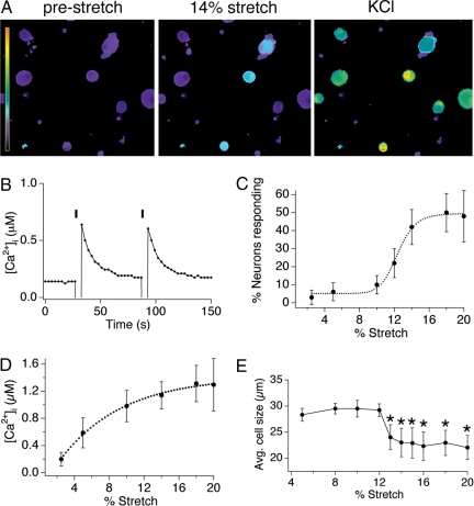
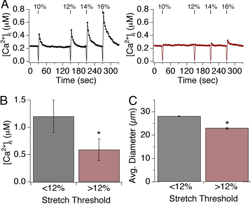
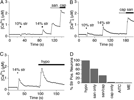
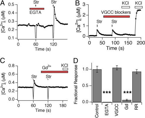
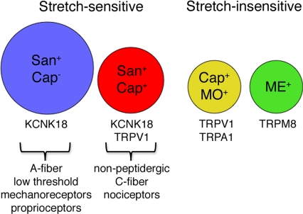
Primary afferent somatosensory neurons mediate our sense of touch in response to changes in ambient pressure. Molecules that detect and transduce thermal stimuli have been recently identified, but mechanisms underlying mechanosensation, particularly in vertebrate organisms, remain enigmatic. Traditionally, mechanically evoked responses in somatosensory neurons have been assessed one cell at a time by recording membrane currents in response to application of focal pressure, suction, or osmotic challenge. Here, we used radial stretch in combination with live-cell calcium imaging to gain a broad overview of mechanosensitive neuronal subpopulations. We found that different stretch intensities activate distinct subsets of sensory neurons as defined by size, molecular markers, or pharmacological attributes. In all subsets, stretch-evoked responses required extracellular calcium, indicating that mechanical force triggers calcium influx. This approach extends the repertoire of stimulus paradigms that can be used to examine mechanotransduction in mammalian sensory neurons, facilitating future physiological and pharmacological studies.


Conflict of interest statement

The authors declare no conflict of interest.

Figures

Fig. 1.
Fig. 1.
Radial stretch activates a subset of trigeminal sensory neurons. (A) Calcium responses of dissociated mouse trigeminal neurons to 14% radial stretch (Middle) and 140 mM KCl ringer's solution (Right). Scale bar indicates the intracellular calcium concentration, ranging from 0.1 to 4 μM [Ca2+]i. (B) Calcium response in a representative cell to two applications of a 14% radial stretch stimulus. (C) Dose–response curve displaying the percentage of neurons activated by varying magnitudes of stretch. (D) Dose–response curve displaying the magnitude of the calcium response triggered by varying magnitudes of stretch. (E) The mean diameter of responding neurons is plotted versus stretch magnitude. For all experiments, n ≥200 cells per point, obtained from a minimum of three different neuronal preparations. All data are reported as means ± SEM.
Fig. 2.
Fig. 2.
Two subsets of sensory neurons display varying sensitivity to stretch. (A) Representative responses of individual neurons to four subsequent applications of radial stretch 10, 12, 14, and 16%. Low-threshold neurons respond to all magnitudes of stretch (black, Left graph), whereas a high-threshold neuron responded only to stretch magnitudes ≥12% (red, Right graph). (B) Low-threshold neurons displayed significantly larger peak calcium transients in response to all levels of stretch versus those observed in high-threshold neurons. (C) Neurons showing sensitivity to low stretch intensity had relatively large soma diameters (28 ± 2.7 μm), whereas those responding only at higher magnitudes were significantly smaller (23 ± 1.4 μm; P < 0.05, one-way ANOVA). Asterisks denotes P < 0.05; n ≥ 10 trials/data point, with ≥30 neurons/trial.
Fig. 3.
Fig. 3.
Radial stretch excites presumptive nociceptors and mechanoreceptors. (A and B) Sensory neurons were stimulated by 10% stretch and 14% stretch stimuli, followed by application of capsaicin (1 μM) and hydroxy-α-sanshool (100 μM), and were analyzed by calcium imaging. Low threshold neurons are insensitive to capsaicin, but display hydroxy-α-sanshool sensitivity. High-threshold neurons are sensitive to both capsaicin and sanshool. (C) Sensory neurons were stimulated by 14% stretch followed by application of 30% hypoosmotic solution (220 mOsm). Both classes of neurons were sensitive to osmotic stimuli. However, only 70% of osmotic-sensitive neurons responded to radial stretch (data not shown). All traces displayed are responses observed in representative cells. (D) Quantitative analysis of concordance between stretch sensitivity and pharmacological attributes. Neurons displaying sensitivity to stretch were examined for activation by capsaicin (Cap; 1 μM), hydroxy-α-sanshool (San; 100 μM), mustard oil (AITC; 100 μM), menthol (ME; 500 μM), or hypotonic (Osmo; 220 mOsm) stimuli. Note the high (>95%) preponderance of stretch sensitivity among sanshool sensitive neurons, as compared with the relatively low (≤2%) concordance between stretch sensitivity and mustard oil or menthol sensitivity.
Fig. 4.
Fig. 4.
Radial stretch activates a calcium-permeable transduction channel that is blocked by gadolinium. (A) Calcium response is shown in a representative cell subjected to 14% stretch in the presence or absence of extracellular EGTA (2 mM). Note lack of response when extracellular calcium was chelated by EGTA. (B) Stretch-evoked calcium transients are independent of voltage-gated calcium channel activity. Calcium response in a representative cell subjected to 14% stretch in the absence and presence of a mixture of voltage-gated calcium channel (VGCC) inhibitors (nifedipine, 30 μM; ω-conotoxin GVIA, 1 μM; ω-conotoxin MVII-C, 10 μM; ω-agatoxin, 250 nM). No response to high (140 mM) extracellular KCl was observed in the presence of these inhibitors, demonstrating bona fide VGCC block. (C) Gadolinium ions inhibit stretch-evoked responses. Calcium response in a representative cell subjected to 14% stretch in the absence or presence of GdCl3 (10 mM). (D) Ruthenium red does not inhibit stretch-evoked calcium responses. Calcium response in a representative cell subjected to 14% stretch in the absence or presence of ruthenium red (20 μM). (E) Quantitative analysis of pharmacological block of stretch-evoked calcium responses. Peak calcium responses to radial stretch in the presence of blockers were normalized to peak obtained in the absence of blockers. Stretch-evoked responses were measure in the presence of vehicle (Control), EGTA (10 mM), GdCl3 (10 mM), or ruthenium red (20 μM). Triple asterisks denote P ≤ 0.001.
Fig. 5.
Fig. 5.
Schematic depicting distinct populations of stretch-sensitive and insensitive neurons. Stretch-sensitive neurons fall into two broad categories: Small-diameter cells (red circle) that are dually sensitive to hydroxy-α-sanshool (San) and capsaicin (Cap), and large-diamater cells (blue circle) that respond to hydroxy-α-sanshool, but not capsaicin. These cells likely correspond to high threshold nociceptors and low threshold proprioceptors, respectively. Stretch-insensitive neurons were predominantly of the small-diameter class and were represented by the subset of capsaicin-sensitive cells that also respond to mustard oil (yellow circle), and a cohort of menthol-sensitive cells (green circle). Circle size depicts relative diameter of the different neuronal subtypes.

References

    1. Garcia-Anoveros J, Corey DP. The molecules of mechanosensation. Annu Rev Neurosci. 1997;20:567–594. - PubMed
    1. Gillespie PG, Walker RG. Molecular basis of mechanosensory transduction. Nature. 2001;413:194–202. - PubMed
    1. Kung C. A possible unifying principle for mechanosensation. Nature. 2005;436:647–654. - PubMed
    1. Lumpkin EA, Caterina MJ. Mechanisms of sensory transduction in the skin. Nature. 2007;445:858–865. - PubMed
    1. Sukharev S, Corey DP. Mechanosensitive channels: Multiplicity of families and gating paradigms. Sci STKE. 2004;2004(219):re4. - PubMed

Publication types