. 2008 Dec 16;105(50):19631-6.
doi: 10.1073/pnas.0809264105. Epub 2008 Dec 5.

Anopheles gambiae males produce and transfer the vitellogenic steroid hormone 20-hydroxyecdysone to females during mating

Affiliations

Anopheles gambiae males produce and transfer the vitellogenic steroid hormone 20-hydroxyecdysone to females during mating

Emilie Pondeville et al. Proc Natl Acad Sci U S A. .

Abstract

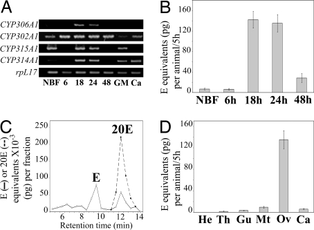
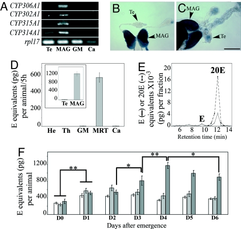
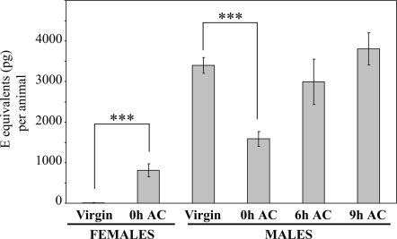
In female insects, the steroid hormone 20-hydroxyecdysone (20E) plays a major role in activating vitellogenesis, a process required for egg development. By contrast with vertebrates, production of large amounts of hormonal steroids has not been reported in adult male insects. In the present study, we analyzed steroidogenesis in both male and female adult of the malaria mosquito Anopheles gambiae and we found that A. gambiae male mosquitoes produce high amounts of the steroid hormone 20E. Importantly, we found that male accessory glands, but not testes, are the source of 20E. Moreover, this steroid hormone is stored in male accessory glands and delivered to females during mating. These findings suggest that male 20E may not act as a true male sex steroid, but more likely as an allohormone. Our results give new insights into species-specific physiological processes that govern the reproductive success of the malaria mosquito. This could thus lead to the identification of new target genes for manipulating male and/or female reproductive success, a promising way to reduce or eliminate mosquito population and therefore to control malaria transmission.


Conflict of interest statement

The authors declare no conflict of interest.

Figures

Fig. 1.
Fig. 1.
Biosynthetic pathway of 20E from cholesterol emphasizing the four terminal steps of steroidogenesis catalyzed by CYP306A1, CYP302A1, CYP315A1, and CYP314A1 (25-, 22-, 2-, and 20-hydroxylases, respectively). (2,22,25dE, 2,22,25-trideoxyecdysone; 2,22dE, 2,22-dideoxyecdysone; 2dE, 2-deoxyecdysone.)
Fig. 2.
Fig. 2.
Ovaries produce 20E after blood meal in A. gambiae. (A) RT-PCR analysis of CYP306A1, CYP302A1, CYP315A1, and CYP314A1 expression pattern in ovaries throughout the first gonotrophic cycle (NBF, ovaries from non-blood-fed females; 6, 18, 24, and 48 h, ovaries at different times PBM) and in different body parts of 24 h PBM blood-fed females (GM, gut and Malpighian tubules; Ca, carcass). (B) In vitro ecdysteroid secretion by ovaries during the first gonotrophic cycle. (NBF, ovaries from non-blood-fed females; 6, 18, 24, and 48 h, ovaries from blood-fed females at different times PBM.) (C) HPLC-EIA analysis of ecdysteroids secreted by ovaries of blood-fed females (24 h PBM). Results are expressed as E (solid line) and 20E (dotted line) equivalents. (D) In vitro ecdysteroid secretion by different body parts of blood-fed females (24 h PBM). (He, head; Th, thorax; Gu and Mt, gut and Malpighian tubules; Ov, ovaries; Ca, carcass.)
Fig. 3.
Fig. 3.
Male accessory glands produce 20E in A. gambiae. (A) RT-PCR analysis of CYP306A1, CYP302A1, CYP315A1, and CYP314A1 expression pattern in different body parts of 6-day-old males. (Te, testes; GM, gut and Malpighian tubules; Ca, carcass.) In this carcass sample, expression level of the control gene RpL17 was lower than in the other tissues examined; replicate experiments confirmed that expression of the steroidogenic genes was at the detection level limit. (B and C) In situ expression pattern of CYP306A1 (B) and CYP314A1 (C) in MRT from 6-day-old males (CYP302A1 and CYP315A1 not shown). (Te, testis; Sb, 250 μm.) (D) In vitro ecdysteroid secretion by different body parts of 6-day-old males. (He, head; Th, thorax; GM, gut and Malpighian tubules; Ca, carcass.) (Inset) In vitro ecdysteroid secretion by separated testes (Te) and MAG of 6-day-old males. (E) HPLC-EIA analysis of ecdysteroids secreted by MRT of 6-day-old males. Results are expressed as E (solid line) and 20E (dotted line) equivalents. (F) Ecdysteroid titers in MRT (white bars), ecdysteroid titers in whole males (light gray bars), and in vitro ecdysteroid secretion by MRT in 5 h of incubation (dark gray bars) from day 0 (D0) to day 6 (D6) PE (*, P < 0.05; **, P < 0.01).
Fig. 4.
Fig. 4.
Males transfer 20E to females during mating. Ecdysteroid titers in virgin females (n = 15), in mated females just after copulation (0 h AC, n = 15), in virgin males (n = 15), in mated males just after copulation (0 h AC, n = 15), and in mated males 6 h (n = 9) and 9 h (n = 12) after copulation (6 h AC; 9 h AC). Ecdysteroids were extracted from each individual mosquito and quantified by EIA. Results are expressed as mean ± SEM in E equivalents (in pg) per animal. Results were subjected to statistical analysis using Mann-Whitney test (***, P < 0.0001).

References

    1. Roll Back Malaria, World Health Organization, UNICEF. World Malaria Report 2005. Geneva: WHO; 2005.
    1. Miller LH, Greenwood B. Malaria–a shadow over Africa. Science. 2002;298:121–122. - PubMed
    1. Coleman PG, Alphey L. Genetic control of vector populations: an imminent prospect. Trop Med Int Health. 2004;9:433–437. - PubMed
    1. Alphey L, et al. Malaria control with genetically manipulated insect vectors. Science. 2002;298:119–121. - PubMed
    1. Catteruccia F. Malaria vector control in the third millennium: progress and perspectives of molecular approaches. Pest Manag Sci. 2007;63:634–40. - PubMed

Publication types

Associated data

LinkOut - more resources