. 2008 Dec 16;105(50):19956-61.
doi: 10.1073/pnas.0810851105. Epub 2008 Dec 5.

In vitro assembly of the Tomato bushy stunt virus replicase requires the host Heat shock protein 70

Affiliations

In vitro assembly of the Tomato bushy stunt virus replicase requires the host Heat shock protein 70

Judit Pogany et al. Proc Natl Acad Sci U S A. .

Abstract

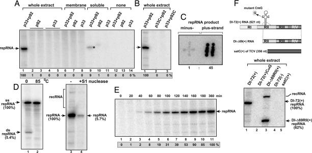
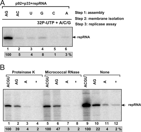
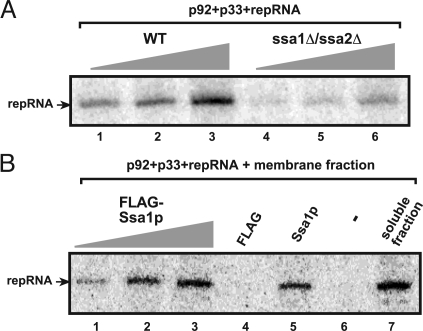
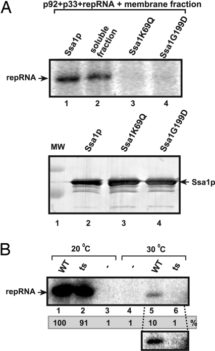
To gain insights into the functions of a viral RNA replicase, we have assembled in vitro and entirely from nonplant sources, a fully functional replicase complex of Tomato bushy stunt virus (TBSV). The formation of the TBSV replicase required two purified recombinant TBSV replication proteins, which were obtained from E. coli, the viral RNA replicon, rATP, rGTP, and a yeast cell-free extract. The in vitro assembly of the replicase took place in the membraneous fraction of the yeast extract, in which the viral replicase-RNA complex became RNase- and proteinase-resistant. The assembly of the replicase complex required the heat shock protein 70 (Hsp70 = yeast Ssa1/2p) present in the soluble fraction of the yeast cell-free extract. The assembled TBSV replicase performed a complete replication cycle, synthesizing RNA complementary to the provided RNA replicon and using the complementary RNA as template to synthesize new TBSV replicon RNA.


Conflict of interest statement

The authors declare no conflict of interest.

Figures

Fig. 1.
Fig. 1.
In vitro assembly of the TBSV replicase. (A) Purified recombinant p33 and p92pol replication proteins of TBSV in combination with DI-72 (+)repRNA were added to the cell-free extract (lanes 1–5), to the membrane fraction (lanes 6–8), to the soluble fraction of the yeast cell-free extract (lanes 9–11) or to the buffer only (lanes 12–14). The denaturing PAGE analysis of the 32P-labeled repRNA products obtained is shown. The full-length repRNA is pointed at by an arrow, whereas the small amount of repRNA product, which is likely due to small level of membrane contamination in the soluble fraction, is marked by an asterisk. (B) Testing the effect of mutations in p33 and p92pol on the activity of the reconstituted TBSV replicase. The denaturing PAGE analysis of the replicase products is as shown in Panel A. Note that p33C lacks the N-terminal portion important for membrane binding, whereas p92C lacks the entire N-terminal portion that overlaps with p33. (C) The 32P-labeled RNA products obtained with the in vitro assembled TBSV replicase programmed with DI-72(+) repRNA were used as probes to hybridize with equal amounts of unlabeled DI-72(+) and (−)RNAs, which had been spotted on the membrane in patterns resembling the shape of “+” and “‖”, respectively. The ratio of (+) and (−) RNA products was calculated by using Imagequant software. (D) Detection of single- and double-stranded RNA products produced by the reconstituted TBSV replicase. (Left) The nondenaturing PAGE shows the dsRNA product after phenol-chloroform extraction (lane 1), which can be fully denatured at 85°C (lane 2). (Right)Denaturing PAGE analysis of the products obtained in the in vitro reconstitution assay without treatment (lane 3) or after S1 nuclease treatment (lane 4), which removes the ssRNA, but not the dsRNA product. recRNA represents recombinant RNAs generated from (+)repRNA (27). (E) Time course analysis of the synthesis of repRNA in the in vitro reconstitution assay. The samples were taken at the shown time points. The denaturing PAGE analysis of the products was done as in A. (F) High template specificity of the reconstituted TBSV replicase in vitro during replication of various viral RNAs. (Top) Schematic representation of the RNA constructs used. Note that satC is a heterologous RNA associated with TCV infections. (Bottom) The denaturing PAGE analysis of the 32P-labeled repRNA products obtained is shown. The head-to-tail recRNA is shown as well.
Fig. 2.
Fig. 2.
Critical role for ribonucleotides during the reconstitution of the TBSV replicase. (A) A stepwise approach was used to separate the possible role of ribonucleotides during the assembly of the TBSV replicase and during RNA synthesis. In step 1, the purified recombinant TBSV p33, p92pol and (+)repRNA were added to the cell-free extract in the presence of various unlabeled ribonucleotides as shown. This was followed by removal of the extra amount of p33, p92pol and repRNA, which were not bound to the membranes of cell-free extract, and then by the standard replicase assay in a buffer containing 32P-UTP and ATP, CTP, and GTP (step 3). The denaturing PAGE analysis of the 32P-labeled repRNA products obtained is shown. (B) The in vitro assembled TBSV replicase forms a protease/ribonuclease-resistant structure in the yeast cell-free extract. The in vitro reconstitution in the presence of various ribonucleotides was done in three steps as described in A. Note that we applied a 15-min treatment with either proteinase K or ribonuclease (micrococcal nuclease) at the end of step 1, before centrifugation (step 2), which removed the proteinase K and the ribonuclease as well as the extra amount of p33, p92pol, and repRNA not bound to the membranes in the cell-free extract. The denaturing PAGE analysis of the 32P-labeled repRNA products obtained is shown.
Fig. 3.
Fig. 3.
Purification and characterization of the in vitro formed TBSV replicase. (A) A schematic presentation of the in vitro assay. The reconstitution assay contained the cell-free extract, the MBP-affinity purified recombinant TBSV p33 and p92pol, various (+)repRNAs as well as various unlabeled ribonucleotides as shown in B. After 1-hour reconstitution, the membrane-bound replicase was solubilized with Triton X-100/SB3–10 detergent, followed by purification on Ni-column of the 6xHis/MBP-tagged p33, which is integral part of the replicase complex. The activity of the affinity-purified TBSV replicase was tested on DI-72(−) RNA added to each sample using the same amount of RNA. (B) The denaturing PAGE analysis of the 32P-labeled DI-72(−) RNA products obtained with the solubilized and purified TBSV replicase is shown. Note that the purified, template-dependent TBSV replicase initiates RNA synthesis precisely from the 3′ end (the product labeled as ti) as well as imprecisely at internal positions on DI-72(−) RNA (indicated as ii products).
Fig. 4.
Fig. 4.
Ssa1p is required for the in vitro assembly of the TBSV replicase. (A) The purified recombinant TBSV p33/p92pol and (+)repRNA were added to the cell-free extract prepared from untransformed (TBSV-free) wt or mutated yeast (ssa1ssa2), which were used in increasing amounts. The denaturing PAGE analysis of the 32P-labeled repRNA products obtained in the in vitro reconstitution assay is shown. (B) Purified recombinant Ssa1p stimulates the assembly of the TBSV replicase in vitro. The membrane fraction of the cell-free extract prepared from untransformed (TBSV-free) wt yeast was programmed with purified recombinant TBSV p33/p92pol and (+)repRNA. The samples also contained 0.2, 1, and 2 μg of Ssa1p (lanes 1–3), FLAG peptide, buffer only (lane 6) or soluble fraction of the cell-free extract as a positive control (lane 7). The 32P-labeled repRNA products obtained in the in vitro reconstitution assay were analyzed with denaturing PAGE.
Fig. 5.
Fig. 5.
Loss-of function Ssa1p mutants cannot stimulate the in vitro assembly of the TBSV replicase. (A) The membrane fraction of the cell-free extract prepared from untransformed (TBSV-free) wt yeast was programmed with purified recombinant TBSV p33 and p92pol and (+)repRNA. The samples also contained purified wt Ssa1p (lane 1), the soluble fraction of the cell-free extract (lane 2), and two loss-of function Ssa1p mutants deficient in ATP hydrolysis (lanes 3 and 4). The 32P-labeled repRNA products obtained in the in vitro reconstitution assay were analyzed with denaturing PAGE. (Bottom) Western blot analysis of affinity-purified Ssa1p and mutants used in the reconstitution assay. (B) Lack of stimulation of the in vitro assembly of the TBSV replicase by a ts mutant of Ssa1p at a nonpermissive temperature. The 32P-labeled repRNA products obtained in the in vitro reconstitution assay were analyzed with denaturing PAGE. The purified wt and ts Ssa1p facilitated the in vitro assembly of the TBSV replicase at 20°C (lanes 1 and 2), whereas only the wt Ssa1p promoted the assembly process at 30°C (lane 5), and the ts mutant was inactive (lane 6). A subset of the panel shows the longer exposure of the portion of the gel.

Similar articles

Cited by

References

    1. Panavas T, Hawkins CM, Panaviene Z, Nagy PD. The role of the p33:p33/p92 interaction domain in RNA replication and intracellular localization of p33 and p92 proteins of Cucumber necrosis tombusvirus. Virology. 2005;338:81–95. - PubMed
    1. Ahlquist P, Noueiry AO, Lee WM, Kushner DB, Dye BT. Host factors in positive-strand RNA virus genome replication. J Virol. 2003;77:8181–88186. - PMC - PubMed
    1. Salonen A, Ahola T, Kaariainen L. Viral RNA replication in association with cellular membranes. Curr Top Microbiol Immunol. 2005;285:139–173. - PMC - PubMed
    1. Nagy PD, Pogany J. Multiple roles of viral replication proteins in plant RNA virus replication. Methods Mol Biol. 2008;451:55–68. - PubMed
    1. Kao CC, Singh P, Ecker DJ. De novo initiation of viral RNA-dependent RNA synthesis. Virology. 2001;287:251–260. - PubMed

Publication types

MeSH terms

LinkOut - more resources