. 2009 Apr;50(4):1692-7.
doi: 10.1167/iovs.08-2375. Epub 2008 Dec 5.

Preferential fluid flow in the human trabecular meshwork near collector channels

Affiliations

Preferential fluid flow in the human trabecular meshwork near collector channels

Cheryl R Hann et al. Invest Ophthalmol Vis Sci. 2009 Apr.

Abstract

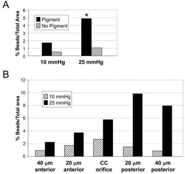
Purpose: To determine whether preferential pathways exist within the human trabecular meshwork, pigmented and nonpigmented regions adjacent to and between collector channels were examined, and the configuration of the juxtacanalicular tissue (JCT) was analyzed.

Methods: Healthy whole human eyes were perfused at 10 or 25 mm Hg with 0.5 mum fluorescent beads. Tissue wedges of pigmented and nonpigmented meshwork (with and without collector channels) were dissected from each eye and examined by confocal microscopy. Bead concentration adjacent to and between collector channels was quantified. The configuration of the JCT adjacent to collector channels from whole eyes perfused at 20 mm Hg was analyzed by light microscopy.

Results: Eyes perfused at 25 mm Hg had more beads adjacent to collector channels in pigmented than in nonpigmented regions (4.9%+/-3.5% vs. 1.1%+/-0.9%; P=0.02). In pigmented regions without collector channels, bead concentration was decreased by fivefold (4.9%+/-3.5% vs. 0.96%+/-0.88%; P=0.04). Perfusion at 25 mm Hg increased beads by threefold under pigmented collector channels compared with the same regions in eyes perfused at 10 mm Hg. Expansion of the JCT occurred more often under collector channels at 25 mm Hg than at 10 mm Hg (44% vs. 17%; P=0.01). The JCT region under collector channels was expanded compared with JCT regions between adjacent collector channels (1053+/-424 microm(2) vs. 571+/-216 microm(2); P<0.001).

Conclusions: Increased levels of beads in pigmented trabecular meshwork adjacent to collector channels suggest preferential flow pathways are present in human trabecular meshwork. At elevated pressure, the JCT region under collector channels is expanded, possibly because of increased fluid flow.


Figures

Figure 1
Figure 1
Dissection of trabecular meshwork (TM) demonstrating pigmentation and collector channels. (A) Tissue wedge of human anterior segment showing variable pigmentation in the trabecular meshwork. Arrows: pigmented areas; arrowheads: nonpigmented areas. (B) Tissue wedge of trabecular meshwork with collector channel (CC) extending from the central region of Schlemm's canal (SC), in characteristic stove-pipe formation, into the sclera (arrows).
Figure 2
Figure 2
Calculation of bead area with the use of imaging software. (A) Total area of the trabecular meshwork was obtained by tracing the inner wall of Schlemm's canal (SC) and the uveal beams as boundaries (yellow). (B) Total area of the fluorescent beads (green channel) was calculated within the total trabecular meshwork area (yellow outline). (C) Regions analyzed included those directly under the collector channel (CC) orifice, those anterior to CC (A1, A2), and those posterior to CC (P1, P2). The bead area in each of these regions was computed as the percentage of total bead area (B, green area). Scale bar, 20 μm.
Figure 3
Figure 3
Analysis of JCT regions under and between collector channels using frontal sections. Images of the expanded JCT region under (A) and between (B) collector channels. JCT demarcation is traced in red. Red arrowheads: center of collector channel orifice (A) and center of analyzed region (B). Area of five consecutive 50-μm regions (R1–R5) was analyzed for area. SC, Schlemm's canal; CC, collector channel. Scale bar, 20 μm.
Figure 4
Figure 4
Comparison of bead density in pigmented and nonpigmented regions of trabecular meshwork. (A) Pigmented regions adjacent to collector channel had higher concentrations of beads than nonpigmented regions in the 10- and 25-mm Hg eyes. *P = 0.02. (B)In 10-mm Hg eyes, the collector channel orifice region had more beads than regions 20 and 40 μm away from the orifice. In the 25-mm Hg, anterior regions moving away from the collector channel orifice contained fewer beads. However, posterior regions contained more beads than the collector channel orifice.
Figure 5
Figure 5
Distribution of beads within expanded JCT regions under collector channels. (A) Confocal volume showing expanded JCT area (arrows) under collector channel (CC) orifice. (B) Green channel of confocal image shown in (A). Fluorescent beads migrated to expanded JCT regions but did not localize to them. Arrows: expanded areas of JCT. Scale bar, 20 μm.
Figure 6
Figure 6
Expanded JCT adjacent to and between collector channels. (AC, red line) JCT boundaries. (A) Collector channel orifice with expanded region on the right side of the micrograph (black arrow). Arrowhead: septum. (B) Region between collector channels showing minimal expansion of the JCT. (C) Expanded JCT under collector channel orifice (black arrows). CC, collector channel; SC, Schlemm's canal. Scale bars, 50 μm.

References

    1. Karg SJ, Garron LK, Feeney ML, McEwen WK. Perfusion of human eyes with latex microspheres. Arch Ophthalmol. 1959;61:68–71. - PubMed
    1. Inomata H, Bill A, Smelser GK. Aqueous humor pathways through the trabecular meshwork and into Schlemm's canal in the cynomolgus monkey (Macaca irus) Am J Ophthalmol. 1972;73:760–789. - PubMed
    1. Tripathi RC. Mechanism of the aqueous outflow across the trabecular wall of Schlemm's canal. Exp Eye Res. 1971;11:116–121. - PubMed
    1. Sabanay I, Gabelt BT, Tian B, Kaufman PL, Geiger B. H7 effects on the structure and fluid conductance of monkey trabecular meshwork. Arch Ophthalmol. 2000;118:955–962. - PubMed
    1. Melamed S, Epstein DL. Alterations of aqueous humour outflow following argon laser trabeculoplasty in monkeys. Br J Ophthalmol. 1987;71:776–781. - PMC - PubMed

Publication types