Comparative Study
. 2009 Jan;12(1):44-52.
doi: 10.1038/nn.2234. Epub 2008 Dec 7.

Stimulation of the insulin/mTOR pathway delays cone death in a mouse model of retinitis pigmentosa

Affiliations
Comparative Study

Stimulation of the insulin/mTOR pathway delays cone death in a mouse model of retinitis pigmentosa

Claudio Punzo et al. Nat Neurosci. 2009 Jan.

Abstract

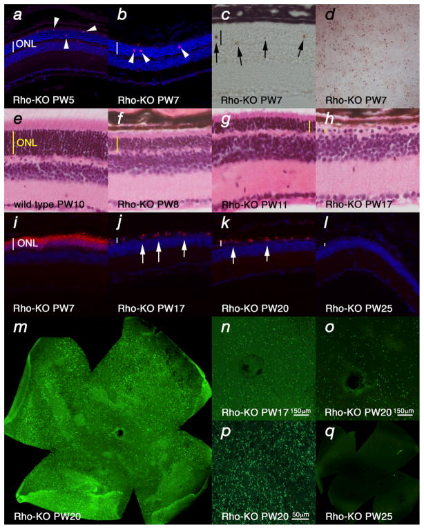
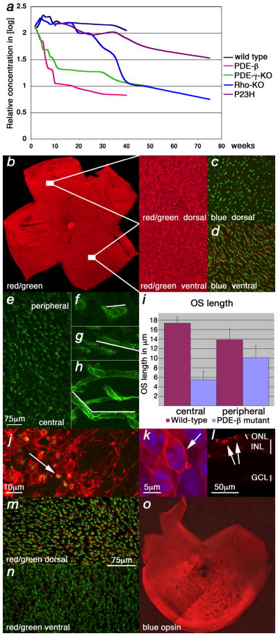
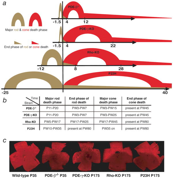
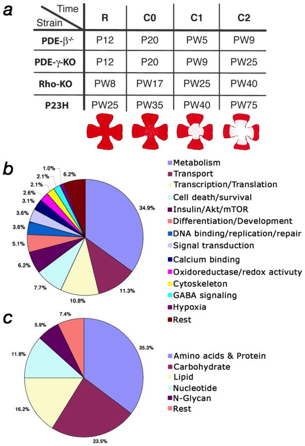
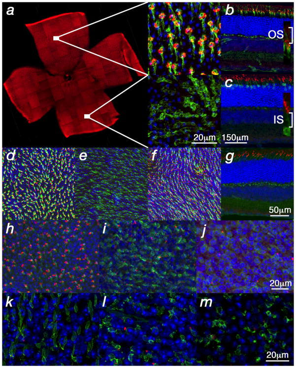
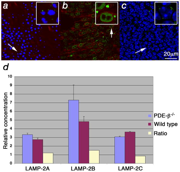
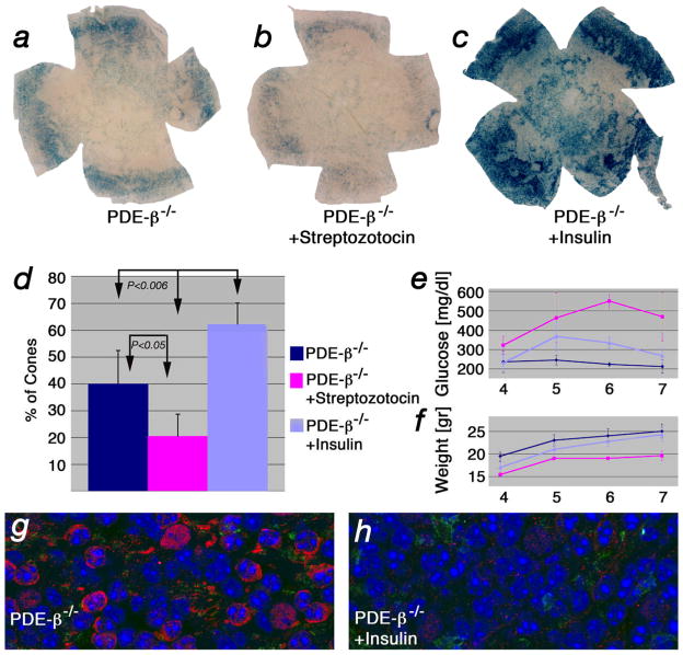
Retinitis pigmentosa is an incurable retinal disease that leads to blindness. One puzzling aspect concerns the progression of the disease. Although most mutations that cause retinitis pigmentosa are in rod photoreceptor-specific genes, cone photoreceptors also die as a result of such mutations. To understand the mechanism of non-autonomous cone death, we analyzed four mouse models harboring mutations in rod-specific genes. We found changes in the insulin/mammalian target of rapamycin pathway that coincided with the activation of autophagy during the period of cone death. We increased or decreased the insulin level and measured the survival of cones in one of the models. Mice that were treated systemically with insulin had prolonged cone survival, whereas depletion of endogenous insulin had the opposite effect. These data suggest that the non-autonomous cone death in retinitis pigmentosa could, at least in part, be a result of the starvation of cones.


Figures

Figure 1
Figure 1
Rod death kinetics in the Rho-KO mutant. (a–d) Onset of rod death seen by cleaved nuclear envelope protein LaminA (a), Cleaved Caspase3 (b) (arrowheads: magenta and red signal respectively) as well as TUNEL (c, d) (arrows: brown signal) (blue in a, b shows nuclear DAPI staining). (d) Shows a retinal flat mount with view onto the photoreceptor layer. (e–h) Progression of rod death determined by the reduction of the ONL as seen by HE staining. (i–q) End phase of rod death assessed by section analysis (i–l) or by retinal flat mounts (m–q). In the Rho-KO the onset of rod death is around PW5 (a) and progresses up to PW25 (l). By PW17 the ONL is reduced to one row of cells (h, j) and in the following 8 weeks the remaining rods die (j–q) as seen by immunofluorescence with an antibody directed against guanine nucleotide protein alpha transducin (Gnat1) on sections of progressively older animals (j–l). (m–q) Retinal flat mounts showing rods visualized by immunofluorescence with an antibody directed against Gnat1. (m) Shows entire retina while (n, o) show higher magnification around the optic nerve head and (p) shows peripheral region. (q) Shows no signal at PW25 where on sections rods were also not detected (l). Age (in postnatal weeks (PW)) is indicated in the panels. Vertical bar in (ac, e–l) indicates thickness of the ONL.
Figure 2
Figure 2
Cone death kinetics. (a) qRT-PCR analysis for Opn1sw during cone degeneration. Changes are in indicated as the logarithm of the relative concentration over time on the Y-axis while X-axis indicates postnatal weeks. (b–h, j, k, m–o) Show retinal flat mounts. (k) Shows a retinal section. Green signal shows PNA expression, red signal shows red/green opsin expression (b, j–n) or blue opsin expression (c, d, o). (b–d) Wild type retina at P35. Red/green opsin (b) and PNA (c, d) expression were detected dorsal and ventral while blue opsin (c, d) was detected only ventrally. (e–g, j–o) Analysis in the PDE-β mutant. (e–g) Central to peripheral gradient of PNA and shortening of cone outer segments (OS). At P20, prior to the major cone death phase, there were fewer elongated OS in the center (e) as compared to the periphery. (f) High magnification of a central or peripheral (g) OS from (e). (h) Wild type OS (white line in f–h marks the OS). (i) Quantification of OS length in central and peripheral regions. The data represents an average of 15 measurements on 3 different retinae of 3 week old mice. With the shortening of OSs during degeneration, red/green opsin was localized throughout the membrane of the cell body and PNA, which detects an extracellular protein(s), was reduced to a small dot attached to the residual OS (j) (arrow: yellow shows red/green and PNA overlap). (k) High magnification of a cone showing red/green localization at the membrane of the main cell body (arrow). (l) Cross section showing red/green in cell body (arrows; j–l P70). Red/ green opsin was detected mainly dorsal (l) during degeneration while PNA (m, n) or blue opsin (o) were not altered (m, n: P21, same scale bar; o: P49).
Figure 3
Figure 3
(a) Schematic representation of the rod and cone death kinetics found in the 4 mouse models of RP. The onset of cone death is set as time zero. The corresponding time windows on the x-axis are given in weeks. For the major rod death phase see text. The major cone death phase was the time until roughly 85% of cones had died. The end phase of cone death was the time thereafter. (b) Summary of rod and cone death kinetics. Time is indicated in postnatal days (P) or postnatal weeks (PW). (c) Immunofluorescence on retinal flat mounts showing the ventral reduction of red/green opsin expression found in the 4 mutants during cone degeneration. Strain and time, in postnatal days, are indicated below each image. (For higher magnification of wild type see figure 2b).
Figure 4
Figure 4
Affymetrix microarray analysis. (a) Equivalent time points in the 4 different mutants at which the microarray analysis was performed (R: approximately halfway through the major phase of rod death; C0: onset of cone death; C1 & C2 first and second time point during cone death respectively). Time is indicated in postnatal days (P) or postnatal weeks (PW). Cartoons depicting the progression of cone death are shown below the corresponding time points. (b) Distribution in percentage of the 195 genes that were annotated. (c) Distribution in percentage of the 68 genes (34.9%) that are part of metabolism in (b).
Figure 5
Figure 5
p*-mTOR in wild type and degenerating retinae. All panels show immunofluorescence on retinal flat mounts (photoreceptor side up) with the exception of (b, c, g) which show retinal sections. Blue shows the nuclear DAPI stain. (a–c) p*-mTOR levels in wild type retinae. (a) Dorsal (up) enrichment of p*-mTOR. Higher magnification of dorsal and ventral region is shown to the right showing p*-mTOR in red and cone segments in green as detected by PNA. (b, c) Dorsal retinal sections stained for p*-mTOR (red signal) and PNA (b) (green signal) or α–β-galactosidase (c) (green signal). The β-galactosidase is under the control of the human red/green opsin promoter and is expressed in all cones (see Material & Methods). The insets in (b, c) show higher magnification of the cone segments suggesting that the p*-mTOR signal is located in the lower part of the outer segment (OS; IS: inner segment). (d–g) Rapamycin treatment of wild type mice leads to downregulation of red/green opsin ventrally (e) but not dorsally (d) (red signal). Ventral blue opsin (f) (red signal) remains unaffected, as does PNA (d–g) (green signal). Rapamycin treatment does also not affect mTOR phosphorylation in wild type (g) (red signal). (h–m) Reduced levels of dorsal p*-mTOR during photoreceptor degeneration (red signal). (h) Wild type control. (i, j) PDE-β mutant. The reduction starts during rod death at P15 (i) as the OSs (green signal: PNA) start to detach from the retinal pigmented epithelium. (i) By P30 only few cones (green signal: α-β-galactosidase) show high levels of p*-mTOR (red signal). (k–l) A similar reduction is seen in dorsal cones of the other three mutants (cones marked in green by PNA). (k) PDE-γ-KO P35. (l) Rho-KO PW20. (m) P23H PW70.
Figure 6
Figure 6
Upregulation of Hif-1α and GLUT1 in cones. All panels show immunofluorescent staining. Left column (a, d, g, h,) shows retinal flat mounts and right column (b, c, e, f, i, j) retinal sections. Blue shows nuclear DAPI staining and green shows cones marked with PNA. (a–f) Staining for HIF-1α (red signal). (a) Wild type (PW10) (inset) showing higher magnification. (b, c) Cross sections in wild type (PW10). (c) DAPI overlap of (b). (d–f) During cone degeneration in PDE-β−/− (PW10) increased levels of HIF-1α are found in cones (d, inset). (e, f) Cross sections show that the increase of Hif-1α occurs mainly in cones (arrows point to cones that at this stage are located within the top layer of the inner nuclear layer). (f) DAPI overlap of (e). (g) GLUT1 expression in wild type (PW10) (red signal). Most of the signal in between the cones reflects expression in rods. (h–j) Increased expression of GLUT1 in cones during degeneration seen in flat mounts (h) and sections (i–j). (i) Overlap of (j) with PNA.
Figure 7
Figure 7
Increased levels of LAMP-2 at the lysosomal membrane. (a–c) Immunofluorescence on retinal flat mounts where LAMP-2 is shown in green, red/green opsin in red and blue signal shows nuclear DAPI stain. Insets in upper right corner (with box) show enlarged cells (arrow). (a) Wild type retinae at PW5 showing lysosome (small green dots) with normal LAMP-2 distribution. Weak red/green opsin signal is detected at the level of the PR nuclei since in wild type it is mainly found in the OSs. (b, c) PDE-β mutant at PW5. (b) Enlarged lysosomes (dots) due to accumulation of LAMP-2 at the lysosomal membrane are seen specifically in cones. (c) Confocal section of same field as in (b) taken at the level of the inner nuclear layer showing levels of LAMP-2 similar to those in wild type (a). (d) qRT-PCR for the 3 different LAMP-2 splice forms showing the relative concentration and the ratios between the PDE-β mutant and wild type.
Figure 8
Figure 8
Insulin levels affect cone survival. (a–c) Retinal flat mounts of PDE-β mutants at PW7 stained for lacZ, to detect cones (see Material & Methods and Supplementary Fig. 8 online). (a) Example of untreated control. (b) Example of mouse injected with streptozotocin. (c) Example of mouse injected daily with insulin. (d) Quantification of cone survival after 4 weeks of treatment. Data represents an average of at least 8 retinae and indicates on the y-axis percentage of cone surface area versus surface area of entire retina (see Supplementary Fig. 9 & 10 online). (e) Measurements of blood glucose levels and body weight (f) performed weekly over the time span of the experiment. (g, h) Immunofluorescent staining on retinal flat mounts for HIF-1α (red signal) and PNA (green signal) in untreated control PDE-β−/− (g) and PDE-β−/− mice treated for 4 weeks with insulin (h). Blue shows nuclear DAPI.

Comment in

References

    1. Madreperla SA, Palmer RW, Massof RW, Finkelstein D. Visual acuity loss in retinitis pigmentosa. Relationship to visual field loss. Arch Ophthalmol. 1990;108:358–61. - PubMed
    1. Steinberg RH. Survival factors in retinal degenerations. Curr Opin Neurobiol. 1994;4:515–24. - PubMed
    1. Mohand-Said S, et al. Normal retina releases a diffusible factor stimulating cone survival in the retinal degeneration mouse. Proc Natl Acad Sci U S A. 1998;95:8357–62. - PMC - PubMed
    1. Streichert LC, Birnbach CD, Reh TA. A diffusible factor from normal retinal cells promotes rod photoreceptor survival in an in vitro model of retinitis pigmentosa. J Neurobiol. 1999;39:475–90. - PubMed
    1. Mohand-Said S, et al. Photoreceptor transplants increase host cone survival in the retinal degeneration (rd) mouse. Ophthalmic Res. 1997;29:290–7. - PubMed

Publication types

MeSH terms