. 2009 Feb 12;28(6):933-6.
doi: 10.1038/onc.2008.445. Epub 2008 Dec 8.

Cancer cells suppress p53 in adjacent fibroblasts

Affiliations

Cancer cells suppress p53 in adjacent fibroblasts

J Bar et al. Oncogene. .

Abstract

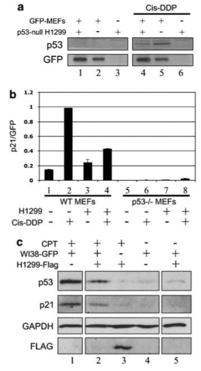
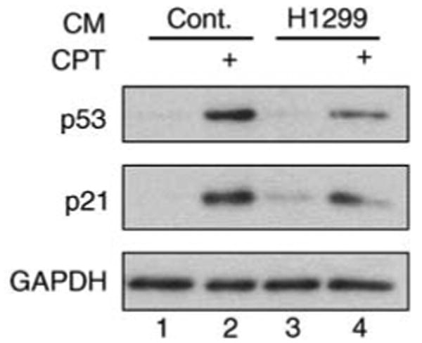
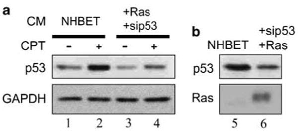
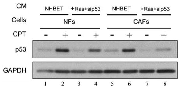
The p53 tumor suppressor serves as a crucial barrier against cancer development. In tumor cells and their progenitors, p53 suppresses cancer in a cell-autonomous manner. However, p53 also possesses non-cell-autonomous activities. For example, p53 of stromal fibroblasts can modulate the spectrum of proteins secreted by these cells, rendering their microenvironment less supportive of the survival and spread of adjacent tumor cells. We now report that epithelial tumor cells can suppress p53 induction in neighboring fibroblasts, an effect reproducible by tumor cell-conditioned medium. The ability to suppress fibroblast p53 activation is acquired by epithelial cells in the course of neoplastic transformation. Specifically, stable transduction of immortalized epithelial cells by mutant H-Ras and p53-specific short inhibitory RNA endows them with the ability to quench fibroblast p53 induction. Importantly, human cancer-associated fibroblasts are more susceptible to this suppression than normal fibroblasts. These findings underscore a mechanism whereby epithelial cancer cells may overcome the non-cell-autonomous tumor suppressor function of p53 in stromal fibroblasts.


Figures

Figure 1
Figure 1
Human cancer cells inhibit the induction of p53 by genotoxic agents in adjacent fibroblasts. (a) Green fluorescent protein (GFP)-expressing mouse embryonic fibroblasts (MEFs) (GFP-MEFs), grown as described (Bar et al., 2004), were plated alone (370 000 cells per 6 cm dish; lanes 2 and 5) or cocultured for 48 h with a twofold excess of p53-null human lung cancer H1299 cells (ATCC; lanes 1 and 4). Cis-DDP (4 μg/ml; Abic, Netanya, Israel) treatment was for 18 h. Cocultures were harvested as is, whereas pure GFP-MEFs control cultures were co-harvested with separately grown, similarly treated cultures of H1299 cells to load similar protein amounts in each lane. Protein was extracted, run on SDS–polyacrylamide gel electrophoresis (PAGE), western blotted for mouse p53 (CM5; Novocastra, Newcastle, UK), and for GFP (clones 7.1 + 13.1; Roche Diagnostics, Mannheim, Germany) as a loading control for MEF-derived proteins. As an additional control, pure cultures of H1299 cells were also similarly processed (lanes 3 and 6). All cell cultures used in this study were routinely tested and found to be mycoplasma free (Logunov et al., 2008). (b) Wild-type (WT) GFP-MEFs (columns 1–4) or p53 knockout GFP-MEFs (columns 5–8) were plated alone or with H1299 cells for 48 h as in (a). Cis-DDP treatment (4 μg/ml) was for 16 h. Total RNA was extracted and p21 mRNA levels determined with mouse-specific primers (s: GGCCCGGAACATCTCAGG, as: AAATCTGTCAGGCTGGTCTGC). Real-time RT-PCR was performed as described (Moskovits et al., 2006). Levels of p21 mRNA were normalized to GFP mRNA (s: GAGCTGAAGGGCATCGACTT, as: CTTGTGCCCCAGGATGTTG). (c) WI-38 fibroblasts stably expressing GFP were cultured alone (3.2 million cells per 10cm dish) or cocultured for 30 h with a threefold access of H1299 cells stably expressing H2B-FLAG CPT (Sigma, Rehovot, Israel; 1 μg/ml) was added for 16 h. GFP-positive cells were fluorescence-activated cell sorting (FACS) sorted either from pure WI-38 cultures (lanes 1 and 4) or from cocultures (lanes 2 and 5). Pure H1299 cultures were collected without GFP gating (lane 3). Extracts loaded correspond to equivalent numbers of cells in each lane. Following SDS–PAGE, western blot analysis was performed for human p53 (mixture of PAb1801 and DO1), p21 (C-19; Santa Cruz Biotechnology, CA, USA), and glyceraldehyde-3-phosphate dehydrogenase (GAPDH) (MAB374; Chemicon International, Chandlers Ford, UK) as a protein loading control. Anti-FLAGantibodies (M2; Sigma-Aldrich, St Louis, MO, USA) were used to confirm the absence of H1299 cells in the fractions collected as pure WI-38 cells.
Figure 2
Figure 2
Cancer cell-conditioned medium inhibits p53 induction. Conditioned medium (CM) was collected from H1299 cells cultured for 48 h in serum-free medium (lanes 3 and 4). Control medium was collected after 48 h incubation without cells (control, lanes 1 and 2). The CM was filtered (0.45 μm) and placed on WI-38 cells, concomitantly with camptothecin treatment (CPT, 1 μg/ml, 19 h) where indicated. Cells were then harvested and subjected to western blot analysis for human p53, p21 (F-5; Santa Cruz Biotechnology) and GAPDH as in Figure 1c.
Figure 3
Figure 3
In vitro-transformed epithelial cells acquire the ability to inhibit p53 in fibroblasts. (a) Normal human bronchial epithelial cells (NHBE; Clonetics-BioWhittaker, San Diego, CA, USA), manipulated to express hTERT either alone (NHBET) or together with mutant H-Ras and p53 short-hairpin RNA (shRNA) (+ Ras + sip53), were generated as described (Milyavsky et al., 2003). Cells were grown with keratinocyte-SFM a serum-free medium supplemented with rEGF and pituitary extract (Gibco Invitrogen Cell Culture, Carlsbad, CA, USA). CM was collected over 48 h with rEGF at 10% the recommended concentration, and placed on WI-38 cells with or without CPT, as in Figure 2. Cells were extracted and western blotted as in Figure 2. (b) Western blot analysis of the epithelial cells, confirming the expression of mutant Ras (C-20; Santa Cruz Biotechnology) and reduction in p53 protein levels by p53 shRNA.
Figure 4
Figure 4
Cancer-associated fibroblasts (CAFs) are more susceptible than normal fibroblasts (NFs) to inhibition of their p53 by CM of transformed epithelial cells. NFs and CAFs were obtained from a surgically resected lung metastasis or from a grossly normal part of the same specimen, of a patient who gave signed informed consent as approved by the Institutional Review Board (IRB). Tissues were cut to small pieces, shaken overnight at 37 °C in collagenase type 4 (250 U/ml; S3J6523; Worthington Biochemical, Lakewood, NJ, USA) in Dulbecco's modified Eagle's medium (DMEM), filtered (100 μm cell strainer; BD Biosciences, San Jose, CA, USA), and plated (DMEM, 20% fetal bovine serum (FBS), 1 mM sodium pyruvate, 2 mM L-glutamine, MEM non-essential amino acid, antibiotics (Beit Haemek, Kibbutz Beit Haemek, Israel) and 60 μM β-mercaptoethanol). After 7–14 days the FBS was reduced to 10%. Fibroblast identity was confirmed by typical morphology, positive vimentin staining and negative cytokeratin staining (data not shown). CM was collected as in Figure 3 from NHBET (lanes 1, 2, 5, 6) and Ras + sip53 (lanes 3, 4, 7, 8) cells, and placed on NFs (lanes 1–4) or CAFs (lanes 5–8) with or without CPT as in Figure 3. Cell extracts were subjected to western blot analysis as in Figure 2.

References

    1. Bar J, Cohen-Noyman E, Geiger B, Oren M. Attenuation of the p53 response to DNA damage by high cell density. Oncogene. 2004;23:2128–2137. - PubMed
    1. Buckbinder L, Talbott R, Velasco-Miguel S, Takenaka I, Faha B, Seizinger BR, et al. Induction of the growth inhibitor IGF-binding protein 3 by p53. Nature. 1995;377:646–649. - PubMed
    1. Campbell IG, Qiu W, Polyak K, Haviv I. Breast-cancer stromal cells with TP53 mutations. N Engl J Med. 2008;358:1634–1635. author reply 1636. - PubMed
    1. Dudley AC, Shih SC, Cliffe AR, Hida K, Klagsbrun M. Attenuated p53 activation in tumour-associated stromal cells accompanies decreased sensitivity to etoposide and vincristine. Br J Cancer. 2008;99:118–125. - PMC - PubMed
    1. Elenbaas B, Weinberg RA. Heterotypic signaling between epithelial tumor cells and fibroblasts in carcinoma formation. Exp Cell Res. 2001;264:169–184. - PubMed

Publication types

MeSH terms

Substances