. 2008;10(6):R104.
doi: 10.1186/bcr2208. Epub 2008 Dec 5.

Buthionine sulfoximine sensitizes antihormone-resistant human breast cancer cells to estrogen-induced apoptosis

Affiliations

Buthionine sulfoximine sensitizes antihormone-resistant human breast cancer cells to estrogen-induced apoptosis

Joan S Lewis-Wambi et al. Breast Cancer Res. 2008.

Erratum in

Abstract

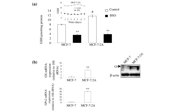
Introduction: Estrogen deprivation using aromatase inhibitors is one of the standard treatments for postmenopausal women with estrogen receptor (ER)-positive breast cancer. However, one of the consequences of prolonged estrogen suppression is acquired drug resistance. Our group is interested in studying antihormone resistance and has previously reported the development of an estrogen deprived human breast cancer cell line, MCF-7:5C, which undergoes apoptosis in the presence of estradiol. In contrast, another estrogen deprived cell line, MCF-7:2A, appears to have elevated levels of glutathione (GSH) and is resistant to estradiol-induced apoptosis. In the present study, we evaluated whether buthionine sulfoximine (BSO), a potent inhibitor of glutathione (GSH) synthesis, is capable of sensitizing antihormone resistant MCF-7:2A cells to estradiol-induced apoptosis.

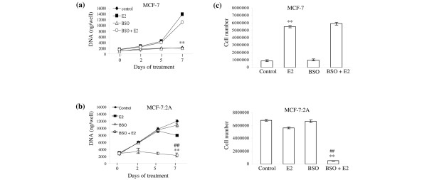
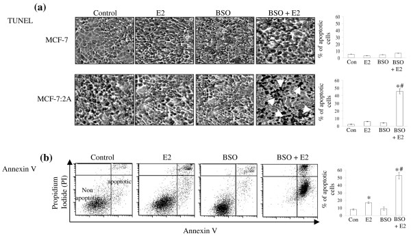
Methods: Estrogen deprived MCF-7:2A cells were treated with 1 nM 17beta-estradiol (E2), 100 microM BSO, or 1 nM E2 + 100 microM BSO combination in vitro, and the effects of these agents on cell growth and apoptosis were evaluated by DNA quantitation assay and annexin V and terminal deoxynucleotidyl transferase dUTP nick end-labeling (TUNEL) staining. The in vitro results of the MCF-7:2A cell line were further confirmed in vivo in a mouse xenograft model.

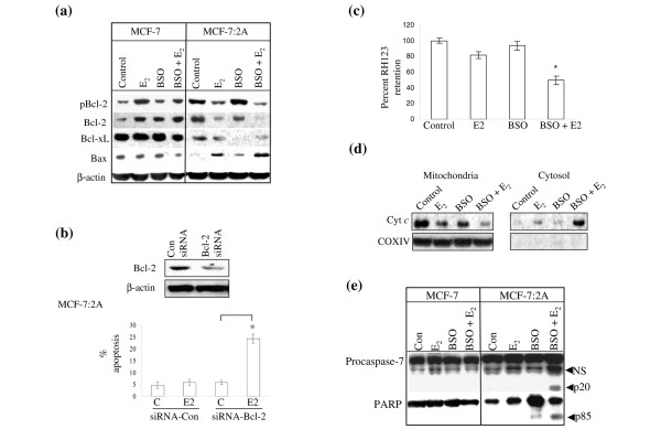
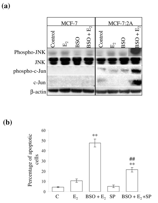
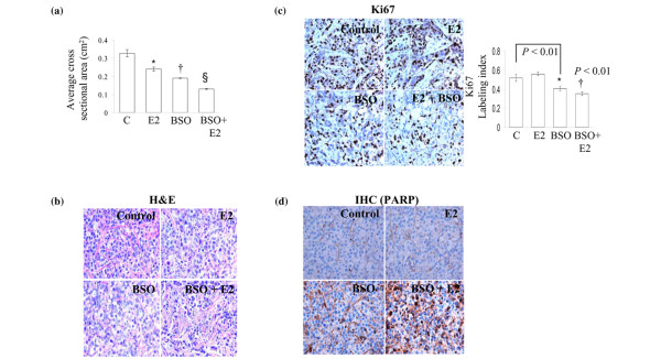
Results: Exposure of MCF-7:2A cells to 1 nM E2 plus 100 microM BSO combination for 48 to 96 h produced a sevenfold increase in apoptosis whereas the individual treatments had no significant effect on growth. Induction of apoptosis by the combination treatment of E2 plus BSO was evidenced by changes in Bcl-2 and Bax expression. The combination treatment also markedly increased phosphorylated c-Jun N-terminal kinase (JNK) levels in MCF-7:2A cells and blockade of the JNK pathway attenuated the apoptotic effect of E2 plus BSO. Our in vitro findings corroborated in vivo data from a mouse xenograft model in which daily administration of BSO either as a single agent or in combination with E2 significantly reduced tumor growth of MCF-7:2A cells.

Conclusions: Our data indicates that GSH participates in retarding apoptosis in antihormone-resistant human breast cancer cells and that depletion of this molecule by BSO may be critical in predisposing resistant cells to E2-induced apoptotic cell death. We suggest that these data may form the basis of improving therapeutic strategies for the treatment of antihormone resistant ER-positive breast cancer.


Figures

Figure 1
Figure 1
Intracellular glutathione (GSH) levels in wild-type MCF-7 cells and antihormone-resistant MCF-7:2A breast cancer cells. (a) MCF-7 and MCF-7:2A cells were seeded at 2 × 10⁶ cells per 100 mm culture plates in phenol red RPMI media containing 10% fetal bovine serum (FBS) and phenol red-free RPMI media containing 10% 4× dextran coated charcoal-treated FBS (SFS), respectively, and after 24 h were treated with nothing (control) (white columns) or 100 μM buthionine sulfoximine (BSO) (black columns) for 24 h. Total cellular glutathione was measured using a Glutathione Colorimetric microplate assay kit, as described in Materials and methods. Columns, mean from three separate experiments; bars, ± standard error of the mean (SEM). **, p < 0.001 compared with control cells; #, p < 0.05 compared with MCF-7 control cells. Insert graph shows glutathione levels in MCF-7:2A cells over a 7-day period. (b) Quantitative real-time polymerase chain reaction (PCR) of glutathione sythetase (GS) (top left) and glutathione peroxidase 2 (GPx2) (bottom left) mRNA expression in MCF-7 and MCF-7:2A cells. **, p < 0.001 compared with MCF-7 control cells. Western blot analysis of GS protein expression in MCF-7:2A cells is also shown (top right).
Figure 2
Figure 2
Effect of buthionine sulfoximine (BSO) plus estradiol on the growth of wild-type MCF-7 cells and antihormone-resistant MCF-7:2A cells. (a) MCF-7 cells were grown in estrogen-free media for 3 days prior to the start of the growth assay. On the day of the experiment, 30,000 cells were seeded in 24-well plates and after 24 h were treated with < 0.1% ethanol vehicle (control), 1 nM 17β-estradiol (E2), 100 μM BSO, or 100 μM BSO plus 1 nM E2 for 7 days. At the indicated time points, cells were harvested and total DNA (ng/well) was quantitated as described in Materials and methods. The data represent the mean of three independent experiments; bars, ± standard error of the mean (SEM). **, p < 0.001 compared with control cells. (b) MCF-7:2A cells were seeded at the same density as MCF-7 cells and were treated similarly. The data represent the mean of three independent experiments; bars, ± SEM. **, p < 0.001 compared with control cells; ##, p < 0.001 compared with estradiol-treated cells. (c) The effect of BSO plus estradiol on cell proliferation was also determined by cell counting using a hemocytometer. For experiment, 0.5 × 10⁶ MCF-7 (top) and MCF-7:2A (bottom) cells were seeded in 15-cm dishes and after 24 h were treated with 1 nM estradiol, 100 μM BSO, or E2 plus BSO combination for 7 days. Data shown represents the mean of three independent experiments; bars, ± SEM. **, p < 0.001 compared with control cells; ##, p < 0.001 compared with estradiol-treated cells.
Figure 3
Figure 3
Buthionine sulfoximine (BSO) plus estradiol induce apoptosis in MCF-7:2A cells. (a) Terminal deoxynucleotidyl transferase-mediated dUTP nick end-labeling (TUNEL) staining for apoptosis in MCF-7:2A cells following BSO plus 17β-estradiol (E2) treatment for 96 h were performed as described in Materials and methods. Slides were photographed through a brightfield microscope under 100 × magnification. TUNEL-positive cells were stained black (white arrows). Columns (right), mean percentage of apoptotic cells (annexin V-positive cells) from three independent experiments performed in triplicate; bars, ± standard error of the mean (SEM). *, p < 0.001 compared with control cells; #, p < 0.001 compared with estradiol-treated cells. (b) Annexin V staining for apoptosis. Cells were seeded in 100 mm plates at a density of 1 × 10⁶ per plate and after 24 h were treated with ethanol vehicle (control), 1 nM E2, or BSO plus E2 for 72 h and then stained with fluorescein isothiocyanate (FITC)-annexin V and propidium iodide (PI) and analyzed by flow cytometry. PI was used as a cell viability marker. Representative cytograms are shown for each group. Quantitation of apoptosis (percentage of control) in the different treatment groups is shown on the right. bars, ± SEM. *, p < 0.05 compared with control cells; #, p < 0.01 compared with estradiol-treated cells.
Figure 4
Figure 4
Effect of buthionine sulfoximine (BSO) and 17β-estradiol (E2) on Bcl-2 family protein expression and mitochondrial function in MCF-7 and MCF-7:2A cells. (a) Western blot analysis for pBcl-2, Bcl-2, Bcl-xL, and Bax protein expression in parental MCF-7 cells and MCF-7:2A cells following 48 h of treatment with ethanol vehicle (Control), 1 nM E2, 100 μM BSO, or E2 + BSO. Equal loading was confirmed by reprobing with an antibody against β-actin. (b) Small interfering RNA (siRNA) knockdown of Bcl-2 partially sensitizes MCF-7:2A cells to E2-induced apoptosis. Cells were transfected with 100 nM siRNA-Bcl-2 or siRNA-Con (control) and expression levels of Bcl-2 was determined by immunoblot analysis (top). Annexin V staining (bottom) showing the effects of siRNA-con and siRNA-Bcl-2 on apoptosis induced by estradiol treatment in MCF-7:2A cells. *, p < 0.001. (c) Loss of mitochondrial potential in MCF-7:2A cells was determined by rhodamine 123 (Rh123) retention assay. The percentage of cells retaining Rh123 in each treatment group was compared with untreated control. (d) Cytochrome c release from the mitochondria to the cytosol after treatment with E2 alone or BSO and E2 for 48 h was determined as described in Materials and methods. Anti-Cox IV antibody was used as a control to demonstrate that mitochondrial protein fractionation was successfully achieved. (e) Cleavage of caspase 7 and poly(ADP-ribose) polymerase (PARP) (72 h) was assessed by western blot using specific antibodies. The upper band of caspase 7 represents the full-length protein and the lower band (p20, arrow) represents the cleaved activated product; NS, nonspecific. Full length PARP is approximately 116 kDa; cleaved (active) PARP is 85 kDa (arrow). The results are representative of three independent experiments.
Figure 5
Figure 5
Activation of c-Jun N-terminal kinase (JNK) signaling pathway in MCF-7:2A cells in response to buthionine sulfoximine (BSO) and 17β-estradiol (E2) treatment. (a) MCF-7 and MCF-7:2A cells were treated with ethanol vehicle (control), 1 nM E2 or 100 μM BSO plus E2 for 48 h and protein levels of phosphorylated JNK, JNK, phosphorylated c-Jun, and c-Jun were analyzed by western blotting. β-Actin was used as a control. (b) Inhibition of JNK activation by SP600125 (SP) partially reverses the apoptotic effect of BSO and estradiol in MCF-7:2A cells. Cells were pretreated with 20 μM SP600125 or vehicle for 24 h, then further incubated for 48 h with 1 nM E2, E2 + 100 μM BSO, 20 μM SP, or E2 + BSO + SP and apoptosis was determined by annexin V-propidium iodide (PI) staining as described in Materials and methods. Columns, mean percentage of apoptotic cells from three independent experiments performed in triplicate; bars, ± standard error of the mean (SEM). **, p < 0.001 compared with control (C) cells; ##, p < 0.01 compared with E2 plus BSO-treated cells.
Figure 6
Figure 6
Buthionine sulfoximine (BSO) inhibits the growth of MCF-7:2A tumors in vivo. Athymic nude mice (4 to 5 weeks old, n = 20) were injected with MCF-7:2A breast cancer cells and after 20 days when tumors had reached a mean cross-sectional area of 0.3 cm2, animals were randomized into 4 groups and were treated with placebo (saline), 17β-estradiol (E2), BSO, or BSO plus E2 for 7 days as described in Materials and methods. BSO (4 mmol/kg weight) was diluted in saline and was injected intraperitoneally daily. (a) Tumor size was measured everyday and cross-sectional area was calculated by multiplying the length (l) by the width (w) by π and dividing the product by 4 (lwπ/4). Data is shown as mean ± standard error of the mean (SEM). *, p < 0.05, control group compared with the E2 group; †, p < 0.002 control group compared with BSO group; § p < 0.001 control group compared with BSO + E2 group. (b) Microscopy of hematoxylin and eosin (H&E)-stained histological sections of MCF-7:2A tumors treated with placebo, E2, BSO, or BSO plus E2. (c) Immunohistochemical analysis of the proliferation marker Ki-67 in MCF-7:2A tumors treated with placebo, E2, BSO, or BSO plus E2. (d) Paraffin-embedded tumor sections of mice treated with E2, BSO, or BSO plus E2 were immunostained for proteolytically cleaved poly(ADP-ribose) polymerase (PARP), which exists only when cells undergo apoptosis. Three to four tumors per treatment group were analyzed.

Similar articles

Cited by

References

    1. Herold CI, Blackwell KL. Aromatase inhibitors for breast cancer: proven efficacy across the spectrum of disease. Clin Breast Cancer. 2008;8:50–64. doi: 10.3816/CBC.2008.n.003. - DOI - PubMed
    1. Santen RJ, Song RX, Masamura S, Yue W, Fan P, Sogon T, Hayashi S, Nakachi K, Eguchi H. Adaptation to estradiol deprivation causes up-regulation of growth factor pathways and hypersensitivity to estradiol in breast cancer cells. Adv Exp Med Biol. 2008;630:19–34. - PMC - PubMed
    1. Jordan VC, O'Malley BW. Selective estrogen-receptor modulators and antihormonal resistance in breast cancer. J Clin Oncol. 2007;25:5815–5824. doi: 10.1200/JCO.2007.11.3886. - DOI - PubMed
    1. Jordan VC. The 38th David A. Karnofsky lecture: the paradoxical actions of estrogen in breast cancer – survival or death? J Clin Oncol. 2008;26:3073–3082. doi: 10.1200/JCO.2008.17.5190. - DOI - PubMed
    1. Santen RJ, Allred DC. The estrogen paradox. Nat Clin Pract Endocrinol Metab. 2007;3:496–497. doi: 10.1038/ncpendmet0535. - DOI - PubMed

Publication types

MeSH terms